. 2009 Jun 15;182(12):7997-8004.
doi: 10.4049/jimmunol.0802884.

Lipopolysaccharide stimulates platelet secretion and potentiates platelet aggregation via TLR4/MyD88 and the cGMP-dependent protein kinase pathway

Affiliations

Lipopolysaccharide stimulates platelet secretion and potentiates platelet aggregation via TLR4/MyD88 and the cGMP-dependent protein kinase pathway

Guoying Zhang et al. J Immunol. .

Abstract

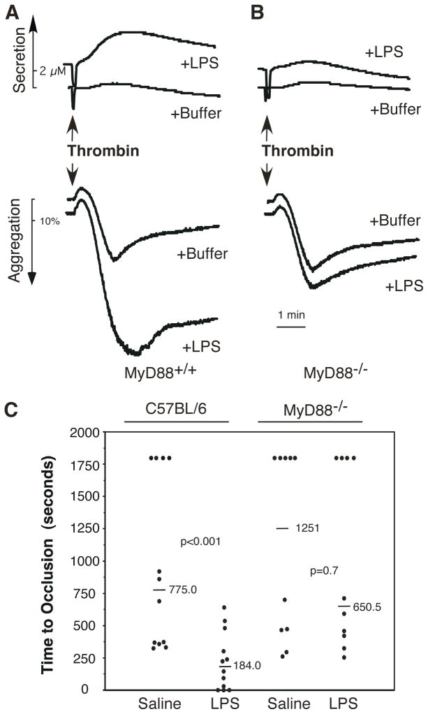
Bacterial LPS induces rapid thrombocytopenia, hypotension, and sepsis. Although growing evidence suggests that platelet activation plays a critical role in LPS-induced thrombocytopenia and tissue damage, the mechanism of LPS-mediated platelet activation is unclear. In this study, we show that LPS stimulates platelet secretion of dense and alpha granules as indicated by ATP release and P-selectin expression, and thus enhances platelet activation induced by low concentrations of platelet agonists. Platelets express components of the LPS receptor-signaling complex, including TLR (TLR4), CD14, MD2, and MyD88, and the effect of LPS on platelet activation was abolished by an anti-TLR4-blocking Ab or TLR4 knockout, suggesting that the effect of LPS on platelet aggregation requires the TLR4 pathway. Furthermore, LPS-potentiated thrombin- and collagen-induced platelet aggregation and FeCl(3)-induced thrombus formation were abolished in MyD88 knockout mice. LPS also induced cGMP elevation and the stimulatory effect of LPS on platelet aggregation was abolished by inhibitors of NO synthase and the cGMP-dependent protein kinase (PKG). LPS-induced cGMP elevation was inhibited by an anti-TLR4 Ab or by TLR4 deficiency, suggesting that activation of the cGMP/protein kinase G pathway by LPS involves the TLR4 pathway. Taken together, our data indicate that LPS stimulates platelet secretion and potentiates platelet aggregation through a TLR4/MyD88- and cGMP/PKG-dependent pathway.


Conflict of interest statement

Disclosures

The authors have no financial conflict of interest.

Figures

Figure 1
Figure 1
LPS potentiates platelet aggregation. A, LPS (0111:B4, 100 μg/ml) was added to washed human platelets and incubated in the aggregometer. B, A subthreshold concentration of thrombin (0.0185 U/ml) was added to washed human platelets immediately followed by addition of various concentrations of LPS (0111:B4) or buffer. C, A subthreshold concentration of collagen (0.3 μg/ml) was added to the washed human platelets followed by addition of LPS (0111:B4, 5 μg/ml) or buffer. D, Quantitative data (mean ± SD) from four experiments described in B (LPS 10 μg/ml) and C. E, A subthreshold concentration of thrombin (0.0185 U/ml) was added to washed human platelets immediately followed by addition of Kdo(2)-Lipid A (10 μg/ml) or buffer. F, Platelet rich-plasma (PRP) from a healthy human donor was added with a collagen (0.6 μg/ml) immediately followed by addition of different sources of LPS (1 μg/ml) or buffer to induce platelet aggregation.
Figure 2
Figure 2
LPS stimulates platelet secretion. A, Washed human platelets were incubated with 1 μg/ml of LPS at 22 °C for 10 min. The release of ATP into the platelet supernatant was determined by luciferin/luciferase assay. B and C, Washed human platelets were incubated with LPS (1 μg/ml) or thrombin (0.025 U/ml) in the presence of a monoclonal anti-human P-selectin antibody, SZ51, for 30 min at room temperature. After washing, platelets were further incubated with a FITC-conjugated anti-mouse IgG. Expression of P-selectin was analyzed by flow cytometry. Quantitative results from three experiments are expressed as the P-selectin expression index (fluorescence intensity (mean) of platelets stimulated with an agonist/fluorescence intensity (mean) of unstimulated platelets) (B). Data from a representative experiment are shown in panel C.
Figure 3
Figure 3
Expression of LPS receptor components in platelets. A, Washed human platelets (1 × 10⁹/ml) were solubilized in 1 × SDS-PAGE sample buffer. TLR4 was detected by immunoblotting with a rabbit anti-human TLR4 polyclonal antibody. β-actin was detected by immunoblotting with a mouse monoclonal antibody against β-actin as a positive control. B, C, and D. Washed human platelets were incubated with a rabbit anti-human TLR4 polyclonal antibody (B), or incubated with a mouse monoclonal antibody, which recognizes the human TLR4/MD2 complex (C), or with a mouse anti-human CD14 monoclonal antibody (D). The platelets were alternatively incubated with rabbit IgG or mouse IgG as a controls. Expression of TLR4, TLR4/MD2 complex, and CD14 was analyzed by flow cytometry. E, Washed human platelets or various concentrations of WBCs from the same donor, were solubilized in 1 × SDS-PAGE sample buffer. MyD88 was detected by immunoblotting with a rabbit anti-human MyD88 polyclonal antibody. β-actin was detected by immunoblotting with a mouse monoclonal antibody against β-actin as a positive control.
Figure 4
Figure 4
LPS potentiates platelet activation via a TLR4-dependent signaling pathway. A, Washed human platelets were pre-incubated with a mouse monoclonal anti-human TLR4 blocking antibody (10 μg/ml) or control mouse IgG at 37°C for 5 min, and then exposed to a subthreshold concentration of thrombin in the presence of LPS (0111:B4, 10 μg/ml) or buffer. B and C, Washed platelets from wild type mice (C57BL/10J) (B) or TLR4 deficient mice (C) were exposed to low dose thrombin in the presence or absence of LPS (0111:B4, 10 μg/ml) to induce platelet secretion and aggregation.
Figure 5
Figure 5
The role of MyD88 in LPS-promoted platelet activation and thrombus formation. A and B, Washed platelets (resuspended in Tyrode’s buffer at 3 × 10⁸/ml) from wild type (A) or MyD88 deficient mice (B) were added with low dose thrombin, followed by addition of LPS (0111:B4, 5 μg/ml) and incubated in the aggregometer. C, FeCl3-induced carotid artery injury was performed and time to occlusive thrombosis recorded as described under Materials and Methods. LPS or saline was injected into fundus oculi of the C57BL/6 or MyD88 deficient mice 1 min after the initiation of carotid artery injury. The occlusion time of each mouse is shown as circles. The bars represent the median occlusion time.
Figure 6
Figure 6
LPS-induced platelet activation involves the cGMP/PKG pathway. A, Washed human platelets were incubated with various concentrations of LPS (0111:B4) at 22 °C for 2 min. cGMP concentrations were determined using a cGMP enzyme immunoassay kit. The reaction was repeated three times using the platelets from the same donor. Results are expressed as mean ± standard error. Statistical significance between groups was determined by t test (p<0.05 for LPS 1 μg/ml, and <0.001 for LPS 10 or 100 μg/ml). The data shown here are representatives of at least three experiments from different donors. B, Washed human platelets were incubated with different sources of LPS (0111:B4 (B4), 055:B5 (B5), or 0127:B8 (B8)) 100 μg/ml at 22 °C for 2 min. C, Washed platelets from TLR4 deficient mice or wild type controls were incubated with LPS (0127:B8) (100 μg/ml) for 0.5 or 2 min. D, Washed human platelets were pre-incubated with mouse IgG, or mouse anti-TLR4 or TLR2 monoclonal antibodies (10 μg/ml) for 5 min at room temperature, and then incubated with LPS (0111:B4) (100 μg/ml) for 2 min. Statistical significance between groups was determined by t test (p<0.05 for TLR4 or TLR2 antibody treated platelets, compared to IgG-treated platelets). E, Washed human platelets were pre-incubated with mouse IgG or an anti-TLR2 blocking antibody for 5 min at 37°C, and then exposed to a low concentration of thrombin in the presence of LPS (0111:B4, 10 μg/ml) or buffer. F, Washed human platelets were pre-incubated with the NOS inhibitor, L-NAME (1 mM), or the PKG inhibitor Rp-pCPT-cGMPS (200 μM), at 37°C for 5 min, and then exposed to a subthreshold concentration of thrombin in the presence of LPS (0111:B4, 10 μg/ml) or buffer to induce platelet aggregation.

References

    1. Tobias PS, Mathison JC, Ulevitch RJ. A family of lipopolysaccharide binding proteins involved in responses to gram-negative sepsis. J Biol Chem. 1988;263:13479–13481. - PubMed
    1. Schumann RR, Leong SR, Flaggs GW, Gray PW, Wright SD, Mathison JC, Tobias PS, Ulevitch RJ. Structure and function of lipopolysaccharide binding protein. Science. 1990;249:1429–1431. - PubMed
    1. Wright SD, Ramos RA, Tobias PS, Ulevitch RJ, Mathison JC. CD14, a receptor for complexes of lipopolysaccharide (LPS) and LPS binding protein. Science. 1990;249:1431–1433. - PubMed
    1. Medzhitov R, Preston-Hurlburt P, Janeway CA., Jr A human homologue of the Drosophila Toll protein signals activation of adaptive immunity. Nature. 1997;388:394–397. - PubMed
    1. Poltorak A, He X, Smirnova I, Liu MY, Van Huffel C, Du X, Birdwell D, Alejos E, Silva M, Galanos C, Freudenberg M, Ricciardi-Castagnoli P, Layton B, Beutler B. Defective LPS signaling in C3H/HeJ and C57BL/10ScCr mice: mutations in Tlr4 gene. Science. 1998;282:2085–2088. - PubMed

Publication types

MeSH terms