. 2009 Jul 8;28(13):1965-77.
doi: 10.1038/emboj.2009.147. Epub 2009 Jun 4.

Molecular recognition of histone lysine methylation by the Polycomb group repressor dSfmbt

Affiliations

Molecular recognition of histone lysine methylation by the Polycomb group repressor dSfmbt

Clemens Grimm et al. EMBO J. .

Abstract

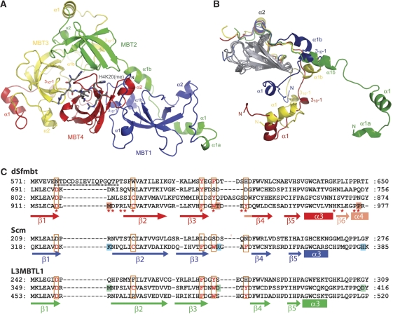
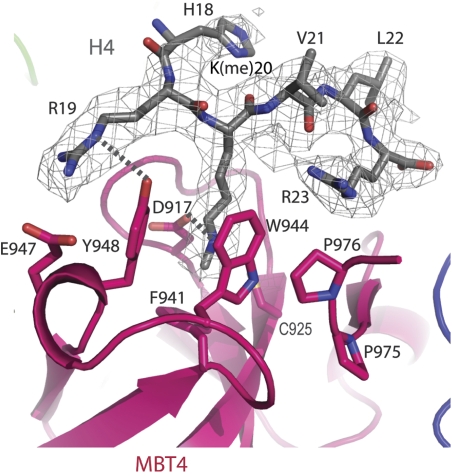
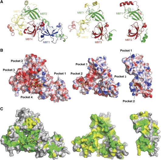
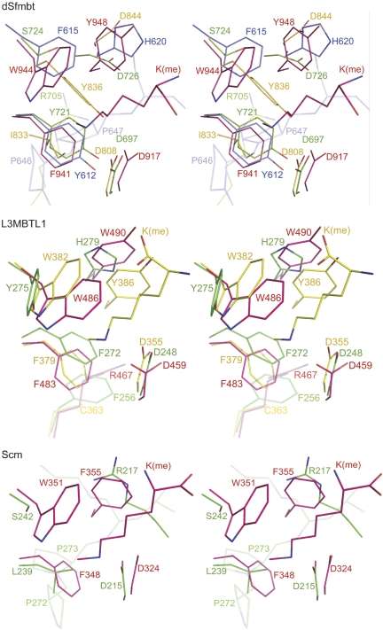
Polycomb group (PcG) proteins repress transcription by modifying chromatin structure in target genes. dSfmbt is a subunit of the Drosophila melanogaster PcG protein complex PhoRC and contains four malignant brain tumour (MBT) repeats involved in the recognition of various mono- and dimethylated histone peptides. Here, we present the crystal structure of the four-MBT-repeat domain of dSfmbt in complex with a mono-methylated histone H4 peptide. Only a single histone peptide binds to the four-MBT-repeat domain. Mutational analyses show high-affinity binding with low peptide sequence selectivity through combinatorial interaction of the methyl-lysine with an aromatic cage and positively charged flanking residues with the surrounding negatively charged surface of the fourth MBT repeat. dSfmbt directly interacts with the PcG protein Scm, a related MBT-repeat protein with similar methyl-lysine binding activity. dSfmbt and Scm co-occupy Polycomb response elements of target genes in Drosophila and they strongly synergize in the repression of these target genes, suggesting that the combined action of these two MBT proteins is crucial for Polycomb silencing.


Figures

Figure 1
Figure 1
Structure of the four MBT-repeat domain of dSfmbt. (A) Ribbon diagram of the four MBT repeats of dSfmbt coloured in blue (repeat 1), green (repeat 2), yellow (repeat 3) and red (repeat 4). Histone H4K20me1 peptide is shown in grey. (B) Superposition of the core folds (grey) of MBT repeats 1–4. For each repeat, helix α2 and the arm regions are coloured according to (A). (C) dSfmbt core domains of repeat 1–4 aligned with core domains of MBT repeats in Drosophila Scm and human L3MBTL1. Positions corresponding to cage-forming residues in dSfmbt repeat 4, Scm repeat 2 and L3MBTL1 repeat 2 are marked with a red box and conserved cage-forming residues are depicted in red. dSfmbt residues contacting the H4K20me1 peptide are indicated with an asterisk. dSfmbt, Scm and L3MBTL1 residues important for differential peptide binding are drawn on red, blue and green background (compare text). N-terminal arm regions are less well conserved and are not included in the alignment.
Figure 2
Figure 2
Methyl-lysine peptide recognition by dSfmbt. Details of the bound histone H4K20me1 peptide binding to the aromatic cage pocket within MBT repeat 4. The simulated annealing omit electron-density map for the ligand is shown in wire–frame mode.
Figure 3
Figure 3
Comparison of the MBT-repeat-domain crystal structures of dSfmbt, L3MBTL1 and Scm. (A) Ribbon diagram of dSfmbt (left), L3MBTL1 (middle) and Scm (right). Equivalent MBT repeats as indicated by comparison of their tertiary structures are depicted with equivalent colours. (B) Electrostatic surface representation of dSfmbt, L3MBTL1 and Scm. The bound peptide ligands are depicted in yellow. In L3MBTL1, the methyl-lysine peptide is bound to MBT repeat 2. (C) Comparison of the surface conservation in dSfmbt, L3MBTL1 and Scm. Conserved regions with >50% sequence conservation are depicted in colour, dark green corresponds to strictly conserved residues. For surface comparison, orthologous sequences were aligned as depicted in Supplementary Figure S2. The following sequences were used for the alignments: dSfmbt: Drosophila melanogaster, Q9VK33, corresponds to the dSfmbt-4MBT crystal structure reported here; Anopheles gambiae, Q7Q0R1; Xenopus laevis, Q32N90; Mus musculus, P59178; Homo sapiens, Q05BQ5; Gallus gallus, Q5ZLC2; Tetraodon nigroviridis, Q4T9N5. L3MBTL1: Homo sapiens, Q9Y468, corresponds to the L3MBTL1 crystal structure (PDB accession code 2RHI); Bos tauru, Q08DF3; Gallus gallus, XP_417302; Danio rerio XP_699604. Scm: Drosophila melanogaster, Q9VHA0, as present in the Scm crystal structure (PDB accession code 2R57); Xenopus tropicalis, Q0IHT6; Homo sapiens, SCML2; Ciona intestinalis Q4H2U6.
Figure 4
Figure 4
Stereo view of superpositons of the MBT-repeat domains of dSfmbt, L3MBTL1 and Scm. Colour code corresponds to Figure 3 with dSfmbt repeats 1, 2, 3 and 4 depicted in blue, green, yellow, and red (top), L3MBTL1 repeats 1, 2 and 3 in green, yellow and red (middle) and Scm repeats 1 and 2 depicted in green and red (bottom).
Figure 5
Figure 5
dSfmbt and Scm co-bind to PREs in PcG target genes. ChIP analysis monitoring dSfmbt and Scm binding in imaginal disc/CNS tissues dissected from wild-type Drosophila larvae. Graphs show the results from three independent immunoprecipitation reactions from different batches of chromatin preparations; ChIP signals were quantified by qPCR and are presented as percentage of input chromatin precipitated at each region, error bars correspond to s.d. The location of PREs (purple boxes) and other regions with respect to transcription start sites in the Ubx, Abd-B, en, ap, Dll, eve and pnr genes are indicated in kilobases; C1–C4 indicate euchromatic and heterochromatic control regions outside these genes (see Supplementary Table 2 for qPCR primer sequences). dSfmbt and Scm proteins are specifically enriched at the PRE of each gene but not at the analyzed intervals in the coding regions of the same genes or in control regions C1–C4.
Figure 6
Figure 6
dSfmbt and Scm interact functionally to maintain Polycomb repression. dSfmbt and Scm act redundantly to maintain repression of Polycomb target genes Abd-B and en in Drosophila. Wing imaginal discs stained with antibodies against Abd-B (red, top) or En protein (red, bottom) as indicated. Left: discs with clones of dSfmbt or calypso single-mutant cells that are marked by the absence of nuclear GFP. Right: disc from ScmD215N-mutant larvae; these animals were trans-heterozygous for ScmD215N and the protein null mutation ScmD1 (Bornemann et al, 1998) and all cells thus express ScmD215N instead of wild-type Scm protein, nuclear GFP was used here to show all nuclei. Middle: ScmD215N/ScmD1 mutant discs with clones of dSfmbt or calypso-mutant cells; the dSfmbt ScmD215N double-mutant and calypso ScmD215N double-mutant cells, respectively, are GFP-negative. Abd-B is not expressed in wild-type wing discs, remains repressed in dSfmbt or calypso single-mutant cells (left, empty arrowheads) or in ScmD215N-mutant discs (right) but is strongly mis-expressed in dSfmbt ScmD215N double-mutant cells (middle, arrowheads). In clones of calypso ScmD215N double-mutant cells (middle), Abd-B is mis-expressed in a small fraction of clone cells (arrowhead) but remains repressed in the majority of clone cells (empty arrowheads). En expression is confined to the posterior-compartment cells of wild-type imaginal discs and this pattern is unchanged in ScmD215N-mutant discs (right); En remains repressed in dSfmbt or in calypso single-mutant clones in the anterior compartment (left, empty arrowheads) with the exception of some dSfmbt-mutant clones in the hinge that show mis-expression of En (filled arrowhead). Note that En is strongly mis-expressed in almost all dSfmbt ScmD215N double-mutant clones in the anterior compartment (middle, arrowheads) but remains repressed in calypso ScmD215N double-mutant clones. Note that only dSfmbt ScmD215N but not calypso ScmD215N double-mutant clones show the tumour-like phenotype (asterisks, see text for details).
Figure 7
Figure 7
Reconstitution of Scm–dSfmbt complexes. (A) FLAG-tagged Scm and untagged dSfmbt, Ph or Pho proteins were affinity purified by FLAG-tag, separated by SDS–PAGE and visualized by Coomassie staining (top). Western blot of corresponding Sf9 total cell-extract input before purification (I) and eluted purified proteins (E) to show relative enrichment of proteins after purification (below). Note that dSfmbt (black arrowhead) forms a stable complex with Flag–Scm (square) whereas Ph (dot) co-purifies less efficiently with Flag–Scm than dSfmbt. Also note that co-expression of Pho with Flag–Scm results in the purification of only Flag–Scm and Pho is not detected by Coomassie staining or western-blot analysis of the eluted purified material. Asterisk marks Flag–Scm degradation products. (B) Immunopurification of different Flag-tagged dSfmbt constructs with full-length Scm (left panel), Flag-tagged Scm constructs with full-length dSfmbt (middle panel) and of Flag-tagged Scm constructs with C-terminally truncated dSfmbt (right panel). Arrowheads and squares indicate dSfmbt and Scm constructs, respectively. Degradation products of Scm protein are indicated by an asterisk. (C) Domain organization of dSfmbt and Scm. Zn-finger domain, MBT repeats and SAM domain are depicted in light grey, grey and black, respectively. Domain borders used for the Scm and dSfmbt constructs are indicated. Brackets indicate the regions of dSfmbt and Scm minimally required for interaction.

Similar articles

Cited by

References

    1. Beuchle D, Struhl G, Muller J (2001) Polycomb group proteins and heritable silencing of Drosophila Hox genes. Development 128: 993–1004 - PubMed
    1. Bornemann D, Miller E, Simon J (1998) Expression and properties of wild-type and mutant forms of the Drosophila sex comb on midleg (SCM) repressor protein. Genetics 150: 675–686 - PMC - PubMed
    1. Celniker SE, Keelan DJ, Lewis EB (1989) The molecular genetics of the bithorax complex of Drosophila: characterization of the products of the Abdominal-B domain. Genes Dev 3: 1424–1436 - PubMed
    1. Cowtan KD, Zhang KY (1999) Density modification for macromolecular phase improvement. Prog Biophys Mol Biol 72: 245–270 - PubMed
    1. de la Fortelle E, Bricogne G (1997) Maximum-likelihood heavy-atom parameter refinement for multiple isomorphous replacement and multiwavelength anomalous diffraction methods. Methods Enzymol 276: 472–494 - PubMed

Publication types

MeSH terms

Associated data