. 2009 Jun 4;4(6):e5798.
doi: 10.1371/journal.pone.0005798.

Mural cell associated VEGF is required for organotypic vessel formation

Affiliations

Mural cell associated VEGF is required for organotypic vessel formation

Lasse Evensen et al. PLoS One. .

Abstract

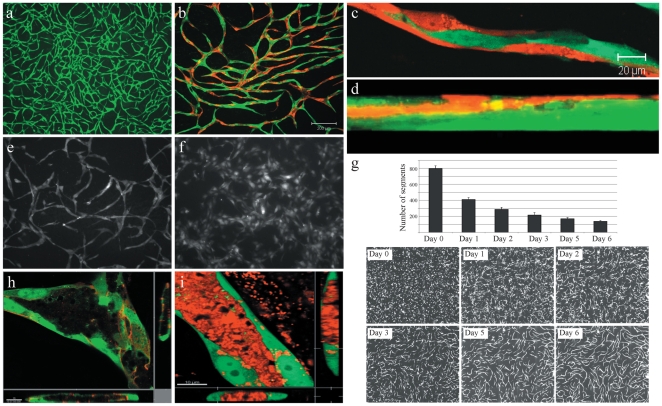
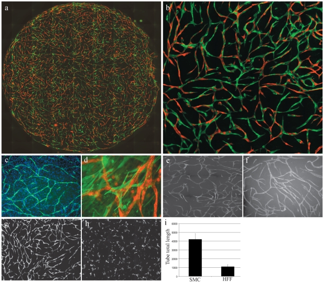
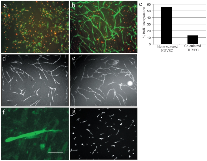
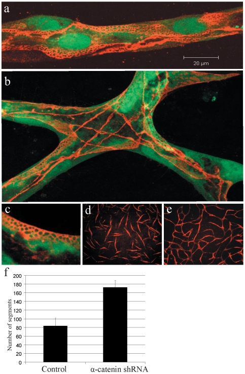
Background: Blood vessels comprise endothelial cells, mural cells (pericytes/vascular smooth muscle cells) and basement membrane. During angiogenesis, mural cells are recruited to sprouting endothelial cells and define a stabilizing context, comprising cell-cell contacts, secreted growth factors and extracellular matrix components, that drives vessel maturation and resistance to anti-angiogenic therapeutics.

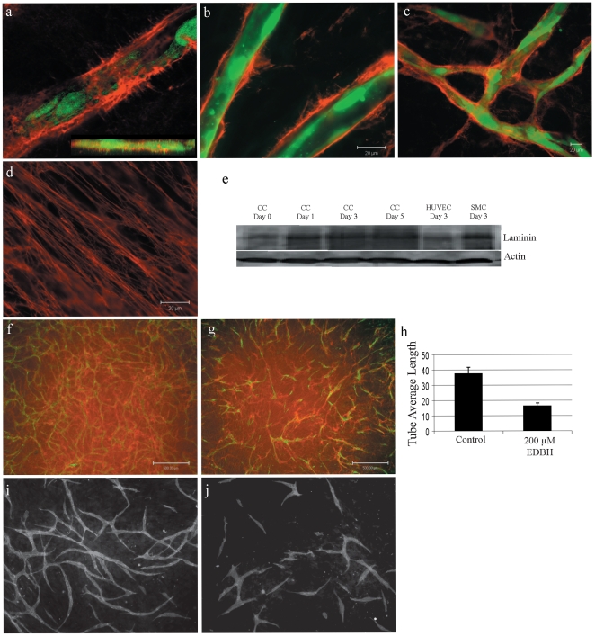
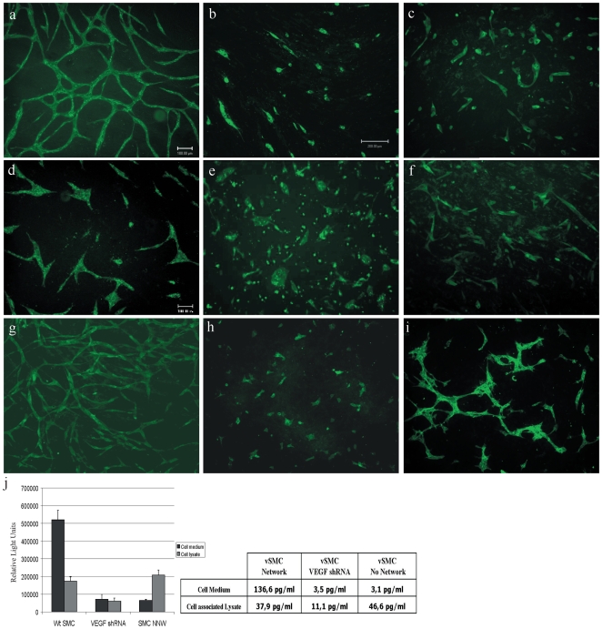
Methods and findings: To better understand the basis for mural cell regulation of angiogenesis, we conducted high content imaging analysis on a microtiter plate format in vitro organotypic blood vessel system comprising primary human endothelial cells co-cultured with primary human mural cells. We show that endothelial cells co-cultured with mural cells undergo an extensive series of phenotypic changes reflective of several facets of blood vessel formation and maturation: Loss of cell proliferation, pathfinding-like cell migration, branching morphogenesis, basement membrane extracellular matrix protein deposition, lumen formation, anastamosis and development of a stabilized capillary-like network. This phenotypic sequence required endothelial-mural cell-cell contact, mural cell-derived VEGF and endothelial VEGFR2 signaling. Inhibiting formation of adherens junctions or basement membrane structures abrogated network formation. Notably, inhibition of mural cell VEGF expression could not be rescued by exogenous VEGF.

Conclusions: These results suggest a unique role for mural cell-associated VEGF in driving vessel formation and maturation.


Conflict of interest statement

Competing Interests: The authors have declared that no competing interests exist.

Figures

Figure 1
Figure 1. Endothelial cells in co-culture with vascular smooth muscle cells generate capillary-like networks.
(A) Live cell fluorescence microscopy of GFP-expressing HUVEC cell capillary-like network (green) after 5 days in co-culture with PA-vSMC (unlabeled). (B) Mixed RFP- and GFP-expressing HUVEC (1∶1) in co-culture with PA-vSMC (unlabeled) at Day 9 demonstrate extensive interaction between ECs within network. (C) Two-color HUVEC networks (9 days) comprise uniform elongated, inter-digitating ECs. (D) Confocal fluorescence microscopy analysis (z-stack) of RFP-expressing HUVEC and GFP-expressing PA-vSMC co-cultures show cells self-organize into distinct layers with ECs residing atop a confluent vSMC layer. (E) Cells grown in co-culture allowing cell-cell contact results in a capillary-like network. (F) Co-cultures in separated by a porous membrane in transwell chambers do not form a network. (G) Temporal image analysis during 6 days of EC network formation shows a decrease in the number of non-networked cells as EC connectivity is established and stabilizes. (H) Lumens form at branch points (inset: z-stacks) and throughout the EC network. (I) TRITC-dextran (10 000 MW) concentrates into patent lumens in Day 9 endothelial networks (inset: z-stacks).
Figure 2
Figure 2. Endothelial- mesenchymal stem cell co-culture generates capillary-like networks.
(A) Live cell fluorescence microscopy whole well image 9×11 montage of RFP-expressing HUVEC - GFP-expressing human dermal microvascular endothelial cell (HuMVEC) – PA-vSMC (unlabeled) tri-culture network at Day 6 (10X objective) (B) Live cell fluorescence microscopy image 9×11montage of interdigitating HUVEC (RFP, red) and HuMVEC (GFP, green) Day 6 tri-culture with vSMC (40×objective). (C) Hoechst nuclear staining of a Day 5 co-culture showing the GFP-expressing endothelial network (green) amid a confluent PA-SMC layer (blue). (D) Confocal microscopy analysis of anti-α-SMA-stained PA-SMC (green) extending filopodia that co-localize with multiple RFP-expressing HUVEC (red) in the vessel-like network. (E) Live cell fluorescence microscopy of GFP-expressing HuMVEC capillary-like network after 5 days in co-culture with PA-vSMC. (F) Live cell fluorescence microscopy of GFP-expressing HUVEC capillary-like network after 5 days in co-culture with primary human bone marrow-derived mesenchymal stem cells (MSC). Comparison of GFP-expressing HUVEC co-cultured with PA-SMC (G) and GFP-expressing HUVEC co-cultured with primary human foreskin fibroblast cells (H). Image analysis shows that HFF are unable to support endothelial network formation (I).
Figure 3
Figure 3. Vascular smooth muscle cells inhibit endothelial cell proliferation and drive Rac1-dependent morphogenesis.
(A) GFP-expressing HUVEC grown in mono-culture exhibit extensive BrdU-incorporation (red). (B) In constrast, co-cultured GFP-expressing HUVEC do not incorporate BrdU, while PA-vSMC SMC divides until confluency. (C) Flow-cytometry analysis showed that HUVEC cells in co-culture have a 4.4-fold decreased proliferation activity compared to a HUVEC mono-culture. Data shown is representative of three individual experiments. (D) Over-expression of the cell cycle inhibitor p21 (GFP-p21) in co-cultured HUVEC did not affect EC network formation relative to GFP-expressing HUVEC control (E). (f) A representative GFP-expressing HUVEC in co-culture at 24 hours post-seeding showing a cell migratory morphology (scalebar = 50 µm). (G) Over-expression of dominant negative Rac1 (GFP-Rac1N17) in co-cultured HUVEC potently inhibited cell morphogenesis and EC network formation.
Figure 4
Figure 4. Endothelial cell networks form adherens junctions.
(A) Adherens junctions visualized by VE-cadherin immunofluorescence (red) delineate the border of interdigititated HUVEC cells (green) in 6 day capillary-like networks. (B) Confocal imaging (collapsed z-stack) of a network branch point where VE-cadherin immunofluorescence defines borders of layered, intersecting endothelial cells. (C) VE-cadherin immunofluorescence showed a lace-like structure. (D) Retroviral vector shRNA-mediated knockdown of α-catenin in co-cultured HUVEC results in inhibition of network formation. Data shown is representative of three independent shRNAs targeting α-catenin. (E) Control shRNA expressing co-cultured HUVECs generate a capillary-like network. (F) Image analysis of fluorescence microscopy images showed a 2-fold increase in distinct HUVEC (segments) detected at 72 hours post co-culture seeding.
Figure 5
Figure 5. Endothelial cell networks are stabilized by basement membrane protein deposition.
(A) Collagen type IV (red) is deposited at GFP-expressing HUVEC cells in co-cultures, completely enveloping the endothelial cells (confocal Z-stack, inset). (B) Collagen type IV and (C) collagen type XVIII have similar EC-associated deposition patterns. (D) SMC-associated laminin expression, 100×objective. (E) Western blot analysis shows that laminin deposition increases during co-culture. (F) Immunofluorescence analysis of collagen IV (red) expression in 72 hour vehicle-treated co-cultures with GFP-expressing HUVEC cells and PA-vSMC (unlabelled). (G) Inhibition of collagen type IV synthesis/maturation by EDBH treatment reduces collagen IV deposition and tube formation. (H) Quantification shows that EDBH-treatment reduces EC network tube average length. (J) Co-cultures incubated in ascorbic acid-deficient medium inhibited EC network formation compared to control (I).
Figure 6
Figure 6. Vascular smooth muscle cell-derived VEGF is required for endothelial capillary-like network formation.
(A) Fluorescence microscopy image of GFP-expressing HUVEC and PA-vSMC co-cultures at day 3 post-seeding. Treatment of EC-vSMC co-cultures with VEGF blocking antibody (Avastin, B) or small molecule VEGFR inhibitor (PTK787/ZK, C) block EC network formation. (D) Transduction of HUVEC with a shRNA targeting VEGFR-2 retroviral vector also strongly inhibited network formation. (E) A human PA-vSMC isolate (NNW) that has very low VEGF production and secretion does not support EC network formation. (F) shRNA-mediated knockdown of VEGF levels in human PA-vSMC inhibits network formation. (G) Overexpression of VEGF165 in PA-vSMC did not affect EC network formation. (H) SV40-immortilized mouse embryonic fibroblasts (SV40-MEF) were unable to drive EC network. (I) HUVEC cells co-cultured with SV40-MEF cells that overexpress VEGF165 form incomplete networks. (J) ELISA quantification of VEGF levels from different vSMC cells.

Similar articles

Cited by

References

    1. Benjamin LE, Golijanin D, Itin A, Pode D, Keshet E. Selective ablation of immature blood vessels in established human tumors follows vascular endothelial growth factor withdrawal. J Clin Invest. 1999;103:159–165. - PMC - PubMed
    1. Bergers G, Hanahan D. Modes of resistance to anti-angiogenic therapy. Nat Rev Cancer. 2008;8:592–603. - PMC - PubMed
    1. Jain RK. Molecular regulation of vessel maturation. Nat Med. 2003;9:685–693. - PubMed
    1. Bergers G, Song S. The role of pericytes in blood-vessel formation and maintenance. Neuro Oncol. 2005;7:452–464. - PMC - PubMed
    1. Adams RH, Alitalo K. Molecular regulation of angiogenesis and lymphangiogenesis. Nat Rev Mol Cell Biol. 2007;8:464–478. - PubMed

Publication types

MeSH terms

Substances