Review
. 2009 Sep;66(17):2851-71.
doi: 10.1007/s00018-009-0050-2. Epub 2009 Jun 4.

Protein complexes in snake venom

Affiliations
Review

Protein complexes in snake venom

R Doley et al. Cell Mol Life Sci. 2009 Sep.

Abstract

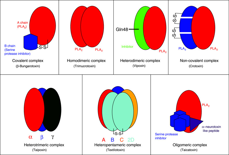
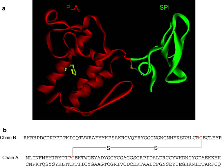
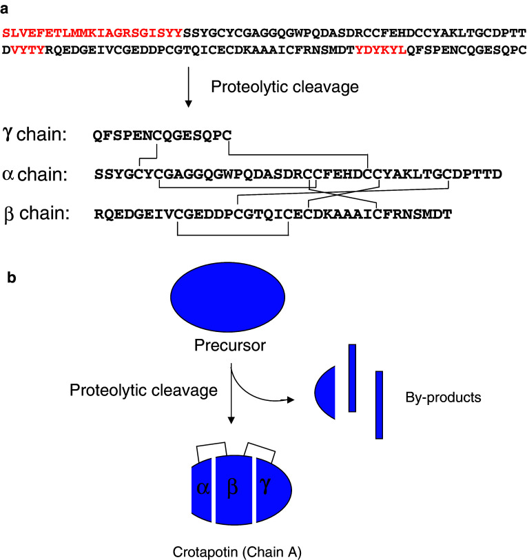
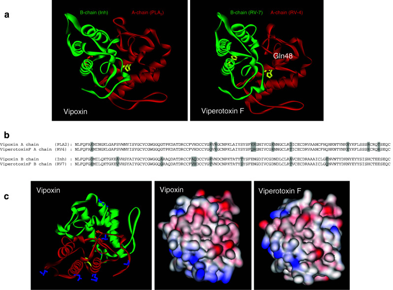
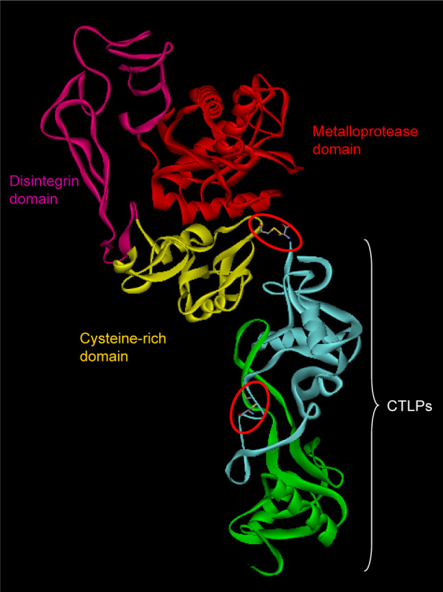
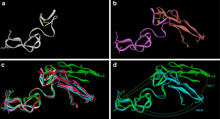
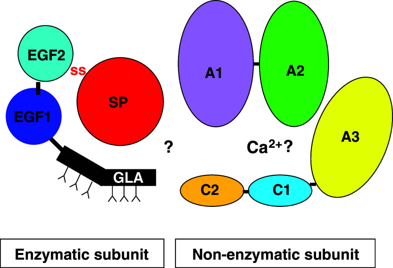
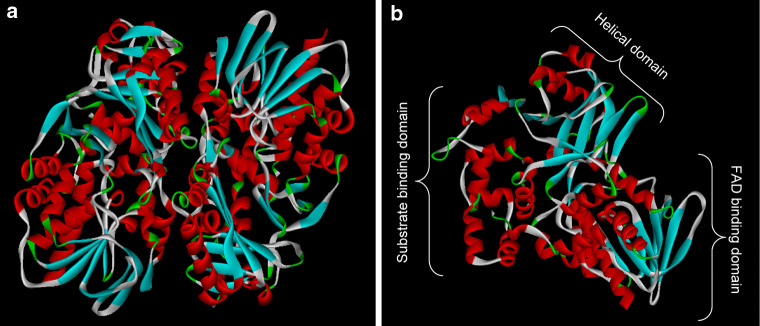
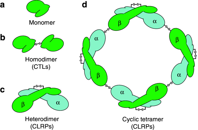
Snake venom contains mixture of bioactive proteins and polypeptides. Most of these proteins and polypeptides exist as monomers, but some of them form complexes in the venom. These complexes exhibit much higher levels of pharmacological activity compared to individual components and play an important role in pathophysiological effects during envenomation. They are formed through covalent and/or non-covalent interactions. The subunits of the complexes are either identical (homodimers) or dissimilar (heterodimers; in some cases subunits belong to different families of proteins). The formation of complexes, at times, eliminates the non-specific binding and enhances the binding to the target molecule. On several occasions, it also leads to recognition of new targets as protein-protein interaction in complexes exposes the critical amino acid residues buried in the monomers. Here, we describe the structure and function of various protein complexes of snake venoms and their role in snake venom toxicity.


Figures

Fig. 1
Fig. 1
Schematic representation of covalent and non-covalent snake venom PLA2 complexes
Fig. 2
Fig. 2
a Ribbon model of β-bungarotoxin (β-BuTx) (PDB ID: 1BUN) showing the disulphide linkage between chain A and chain B. b Inter-chain disulphide bridge is formed between (highlighted in red) 1st Cys of chain A and 6th Cys of chain B, and the linkage is shown in both the ribbon model and amino sequences
Fig. 3
Fig. 3
a Proteolytic cleavage of chain A precursor of crotoxin. Chain A precursor undergoes proteolytic cleavage to form three polypeptide α, β and γ chains. These polypeptides are linked by disulphide bridges. The inter-chain disulphide bridge is shown in straight lines. b Schematic representation of proteolysis and inter-chain disulphide bridge
Fig. 4
Fig. 4
a Ribbon model of vipoxin (PDB ID: 1AOK) and viperotoxin F (PDB ID: 1OQS) showing PLA2 subunit (red) and its inhibitor (green). The active site residue, His48, is replaced with Gln48 in the inhibitor, which is shown in a stick and ball model. b Alignment of amino acid sequence of chain A and B of vipoxin and viperotoxin F. The substituted amino acid residues are highlighted in grey. c The surface residues that are substituted in vipoxin are shown in a stick model (blue color). The surface charge of vipoxin and viperotoxin F is shown
Fig. 5
Fig. 5
Ribbon model of RVV-X (PDB ID: 2E3X) showing different domains: metalloproteinase domain (red), disintegrin domain (megneta), cysteine rich domain (yellow) and CTLP domain (green and turquoise). The interchain disulphide bridges are encircled (red)
Fig. 6
Fig. 6
Dimeric disintegrins. (a) Disintegrin monomer (PDB ID: 1J2L) (white ribbon). RGD is shown as a white stick model. Intramolecular disulfide bond (C6:C14) is shown in the yellow stick. b Homodimeric disinegrin (PDB ID: 1RMR) (pink and orange ribbons). Side chains of RGD are shown as pink/orange sticks. Intermolecular disulfide bonds (C7:C12) are shown in the yellow stick. c Superimposition of homodimeric disintegrins (PDB ID: 1RMR in pink ribbon, PDB ID: 1TEJ in cyan ribbon) and heterodimeric integrins (PDB ID: 1Z1X in red ribbon, PDB ID: 3C05 in green ribbon) on monomeric disintegrin (PDB ID: 1J2L in white ribbon). RGDs are shown as the white stick in 1J2L, as the pink stick in 1RMR, as the cyan stick in 1TEJ and as the green stick in 3C05. MLD is shown as the red stick in 1Z1X. d For 1RMR, the angle between chain A Gly43 Cα, chain B Cys6 Cα, and chain B Gly43 Cα is 100.8°; for 1Z1X, the angle between chain A Gly44 Cα, chain B Cys7 Cα and chain B Gly44 Cα is 136.1°
Fig. 7
Fig. 7
Diagrammatic representation of Pseutarin C. The enzymatic subunit contains the Gla, EGF1, EGF2 and serine protease domain, whereas the enzymatic subunit contains the A1–A3 and C1–C2 domain. The Gla and EGF domains are linked to the serine protease domain by a disulphide bridge. The glycosylation sites of the Gla domain are shown with “Y” in the Figure. The non-enzymatic subunit contains a heavy chain and a light chain similar to mammalian factor Va. The heavy chain contains A1 and A2 domains, whereas light chain contains the A3, C1 and C2 domains. The interaction between the enzymatic and non-enzymatic subunit is not yet know and hence shown with a “?” symbol
Fig. 8
Fig. 8
Ribbon model of l-amino acid oxidase (PDB ID: 1F8S) showing the homodimer (a) and monomer (b). The monomer has three different domains, the FAD-binding domain, helical domain and substrate-binding domain, which are shown in the ribbon model
Fig. 9
Fig. 9
Schematic representation of C-type and C-type lectin-like proteins from snake venom
Fig. 10
Fig. 10
Ribbon model of rattlesnake venom lectins (RSL) (PDB ID: 1JZN) showing pentameric structure
Fig. 11
Fig. 11
a, b Ribbon and schematic model of rattlesnake venom lectins (RSL) monomer (PDB ID: 1JZN) and factor IX/X-binding protein from Trimeresurus flavoviridis (PDB ID: 1IXX). The amino acid residues involved in domain swapping are shown in the stick model. The ligand-binding site in IX/X-binding protein formed by domain swapping is shown in the dotted arc. c Alignment of amino acid sequence of chain A of RSL, chain A and B of IX/X binding protein. The amino acid residues involved in domain swapping are highlighted in yellow color. The extra cysteine residues that form the interchain disulphide bridge in IX/X binding between chain A and B are boxed and marked (arrowhead)
Fig. 12
Fig. 12
Three-finger toxin (3FTxs) complexes. Schematic and ribbon model of κ-bungarotoxin (non-covalent) (a) and iriditoxin (covalent) (b). The interchain disulphide bridge of irditoxin is encircled

Similar articles

Cited by

References

    1. Slotta K, Fraenkel-Conrat H. Schlangengifte-III. Mitteilung: Reinigung und Krystallisation des Klapperschlangen-Giftes. Ber Dtsch Chem Ges. 1938;71:1076–1081. doi: 10.1002/cber.19380710527. - DOI
    1. Kini RM. Excitement ahead: structure, function and mechanism of snake venom phospholipase A2 enzymes. Toxicon. 2003;42:827–840. doi: 10.1016/j.toxicon.2003.11.002. - DOI - PubMed
    1. Bon C. Multicomponent neurotoxic phospholipases A2. In: Kini RM, editor. Phospholipase A2 enzyme: structure, function and mechanism. Chichester: Wiley; 1997. pp. 269–285.
    1. Chang CC, Lee CY. Isolation of neurotoxins from the venom of Bungarus multicinctus and their modes of neuromuscular blocking action. Arch Int Pharmacodyn Ther. 1963;144:241–257. - PubMed
    1. Chu CC, Li SH, Chen YH. Resolution of isotoxins in the beta-bungarotoxin family. J Chromatogr A. 1995;694:492–497. doi: 10.1016/0021-9673(94)01173-C. - DOI - PubMed

Publication types

MeSH terms

LinkOut - more resources