

doi: 10.1007/s00249-009-0472-7.
Epub 2009 Jun 3.
An ultrasoft X-ray multi-microbeam irradiation system for studies of DNA damage responses by fixed- and live-cell fluorescence microscopy
Affiliations
- PMID: 19495740
- PMCID: PMC2701496
- DOI: 10.1007/s00249-009-0472-7
Item in Clipboard
An ultrasoft X-ray multi-microbeam irradiation system for studies of DNA damage responses by fixed- and live-cell fluorescence microscopy
Eur Biophys J.
2009 Jul.
Abstract
Localized induction of DNA damage is a valuable tool for studying cellular DNA damage responses. In recent decades, methods have been developed to generate DNA damage using radiation of various types, including photons and charged particles. Here we describe a simple ultrasoft X-ray multi-microbeam system for high dose-rate, localized induction of DNA strand breaks in cells at spatially and geometrically adjustable sites. Our system can be combined with fixed- and live-cell microscopy to study responses of cells to DNA damage.
Figures
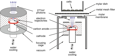
Schematic representation of the multi-microbeam irradiation setup
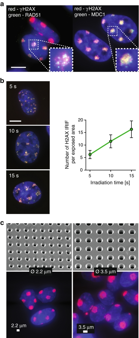
Induction of DNA double-strand breaks by multi-microbeam irradiation. a MMB irradiation induces DSB responses. U2OS cells were irradiated for 6 min through a metal mesh filter with openings of 2.2 μm in diameter, fixed 30 min later, and stained for DNA (blue), γH2AX (red), and Rad51 (green, left panel) or MDC1 (green, right panel). Inset shows magnification of a single irradiated area. Scale bar 10 μm. b The number of DSBs induced by MMB irradiation depends linearly on irradiation time. Left panels show U2OS cells irradiated for 5, 10, or 15 s through a metal mesh filter with openings of 2.2 μm in diameter, fixed 5 min later, and stained for DNA (blue), γH2AX (red), and MDC1 (green). Right panel shows numbers of γH2AX foci representing individual DSBs per exposed area in cells from b. c The geometry of exposed areas can be controlled by applying metal mesh filters of various parameters. Cells were irradiated for 6 min using filters with openings of 2.2 μm (left) or 3.5 μm in diameter (right), fixed 5 min later, and stained for DNA (blue) and γH2AX (red). Upper panels show mesh filters used, imaged by a scanning electron microscope. Lower panels show stained cells irradiated using the respective filters. Scale bar 2.2 μm (left panel) and 3.5 μm (right panel)
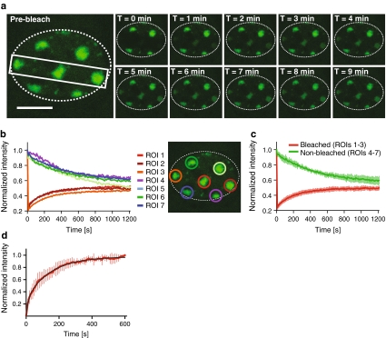
Dynamics of 53BP1-GFP at areas of DNA damage induced by the MMB system. a U2OS cells expressing 53BP1-GFP were irradiated for 6 min through a metal mesh filter with openings of 2.2 μm in diameter and mounted under a confocal microscope. The indicated area of the selected cell was then bleached, and the cell was imaged for 20 min at intervals of 10 s. The gallery shows images captured at indicated times after bleaching. The perimeter of the cell nucleus is indicated by the dotted line. Scale bar 10 μm. b Quantification of the fluorescence intensities measured at the different ROIs in the cell (a) at indicated times after bleaching. The fluorescence intensities at each ROI were normalized to the intensities at the respective ROI before bleaching. c Quantification of average fluorescence intensities at bleached (1–3) and non-bleached (4–7) regions at indicated times after bleaching. Error bars indicate standard deviations. d Quantification of recovery of 53BP1-GFP intensity at damaged chromatin areas after photobleaching. The graph represents normalized average intensity of the bleached areas obtained from measurement of 10 cells. The black curve represents nonlinear fit obtained as described earlier (Bekker-Jensen et al. 2005). Error bars represent standard deviation
References
-
- {'text': '', 'ref_index': 1, 'ids': [{'type': 'DOI', 'value': '10.1088/0305-4608/11/6/014', 'is_inner': False, 'url': 'https://doi.org/10.1088/0305-4608/11/6/014'}]}
- Agarwal BK, Sparrow JH (1981) Line intensities in the soft X-ray region. J Phys F Met Phys 11:1303–1309. doi:10.1088/0305-4608/11/6/014
-
- {'text': '', 'ref_index': 1, 'ids': [{'type': 'DOI', 'value': '10.1083/jcb.200503043', 'is_inner': False, 'url': 'https://doi.org/10.1083/jcb.200503043'}, {'type': 'PMC', 'value': 'PMC2171401', 'is_inner': False, 'url': 'https://pmc.ncbi.nlm.nih.gov/articles/PMC2171401/'}, {'type': 'PubMed', 'value': '16009723', 'is_inner': True, 'url': 'https://pubmed.ncbi.nlm.nih.gov/16009723/'}]}
- Bekker-Jensen S, Lukas C, Melander F, Bartek J, Lukas J (2005) Dynamic assembly and sustained retention of 53BP1 at the sites of DNA damage are controlled by Mdc1/NFBD1. J Cell Biol 170:201–211. doi:10.1083/jcb.200503043 - PMC - PubMed
-
- {'text': '', 'ref_index': 1, 'ids': [{'type': 'DOI', 'value': '10.1083/jcb.200510130', 'is_inner': False, 'url': 'https://doi.org/10.1083/jcb.200510130'}, {'type': 'PMC', 'value': 'PMC2063811', 'is_inner': False, 'url': 'https://pmc.ncbi.nlm.nih.gov/articles/PMC2063811/'}, {'type': 'PubMed', 'value': '16618811', 'is_inner': True, 'url': 'https://pubmed.ncbi.nlm.nih.gov/16618811/'}]}
- Bekker-Jensen S, Lukas C, Kitagawa R, Melander F, Kastan MB, Bartek J et al (2006) Spatial organization of the mammalian genome surveillance machinery in response to DNA strand breaks. J Cell Biol 173:195–206. doi:10.1083/jcb.200510130 - PMC - PubMed
-
- {'text': '', 'ref_index': 1, 'ids': [{'type': 'DOI', 'value': '10.1093/rpd/ncl508', 'is_inner': False, 'url': 'https://doi.org/10.1093/rpd/ncl508'}, {'type': 'PubMed', 'value': '17327240', 'is_inner': True, 'url': 'https://pubmed.ncbi.nlm.nih.gov/17327240/'}]}
- Chang S, Zhang J, Bordelon D, Schreiber E, Cox A, Zhou O (2006) Development of a nanotechnology based low-LET multi-microbeam array single cell irradiation system. Radiat Prot Dosimetry 122:323–326. doi:10.1093/rpd/ncl508 - PubMed
-
- {'text': '', 'ref_index': 1, 'ids': [{'type': 'PubMed', 'value': '7335101', 'is_inner': True, 'url': 'https://pubmed.ncbi.nlm.nih.gov/7335101/'}]}
- Cremer C, Cremer T, Zorn C, Zimmer J (1981) Induction of chromosome shattering by ultraviolet irradiation and caffeine: comparison of whole-cell and partial-cell irradiation. Mutat Res 84:331–348. doi:10.1016/0027-5107(81)90202-5 - PubMed
Publication types
MeSH terms
Substances
LinkOut - more resources
Full Text Sources
Medical
