. 2009 Jun 4:6:56.
doi: 10.1186/1742-4690-6-56.

Intracellular interactions between APOBEC3G, RNA, and HIV-1 Gag: APOBEC3G multimerization is dependent on its association with RNA

Affiliations

Intracellular interactions between APOBEC3G, RNA, and HIV-1 Gag: APOBEC3G multimerization is dependent on its association with RNA

Yeshitila N Friew et al. Retrovirology. .

Abstract

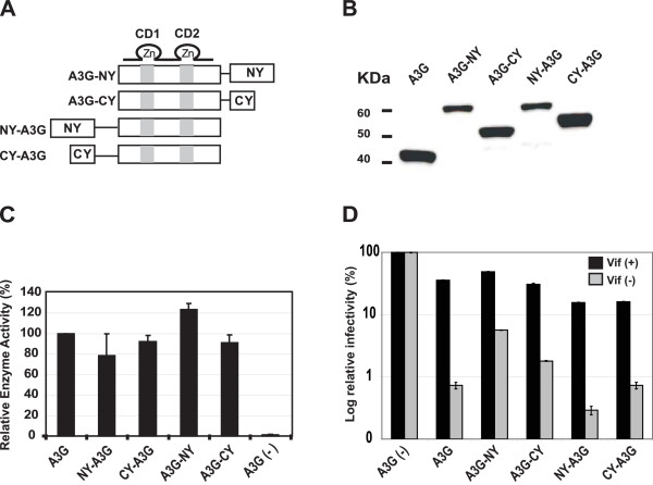
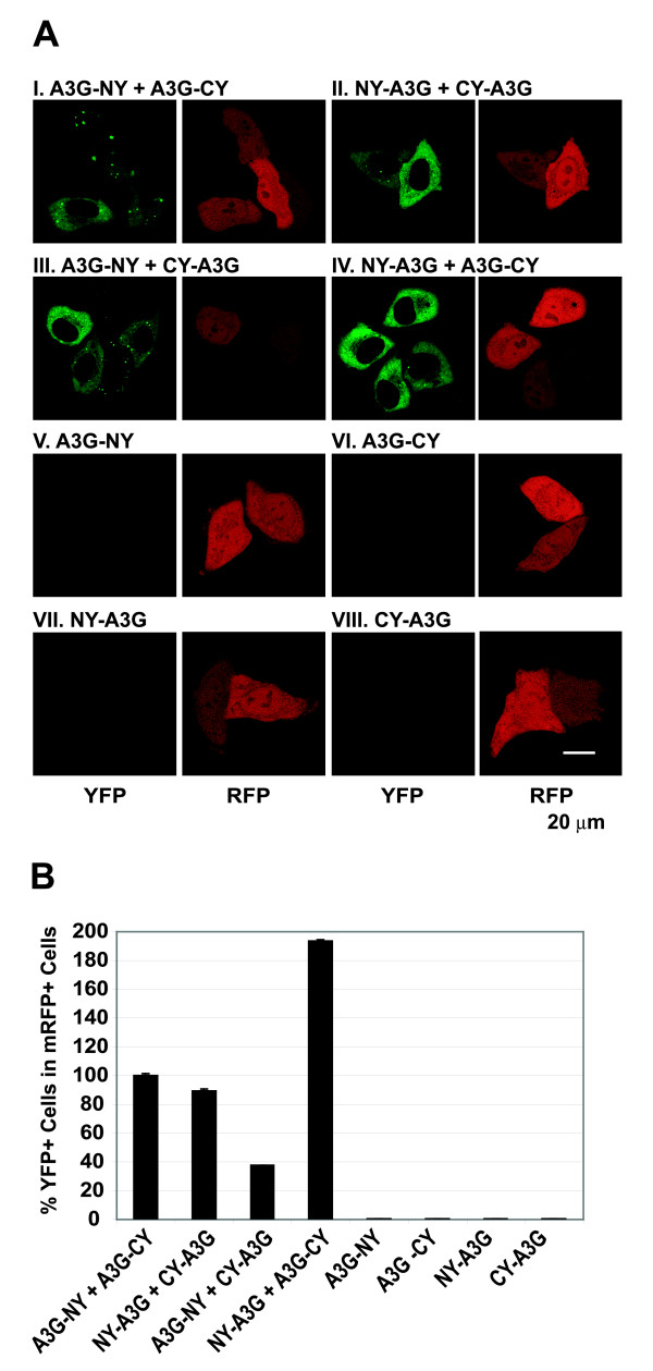
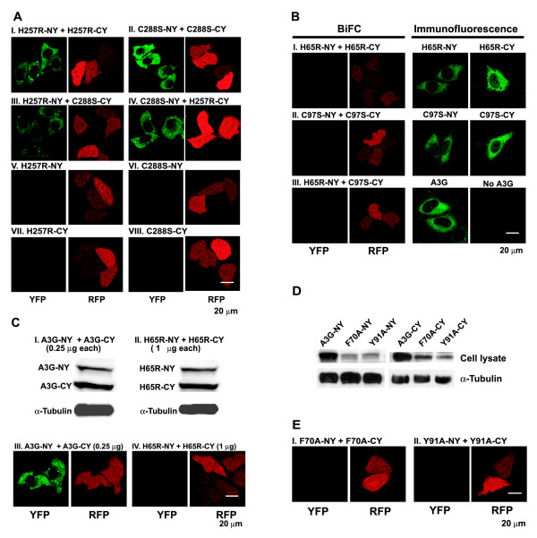
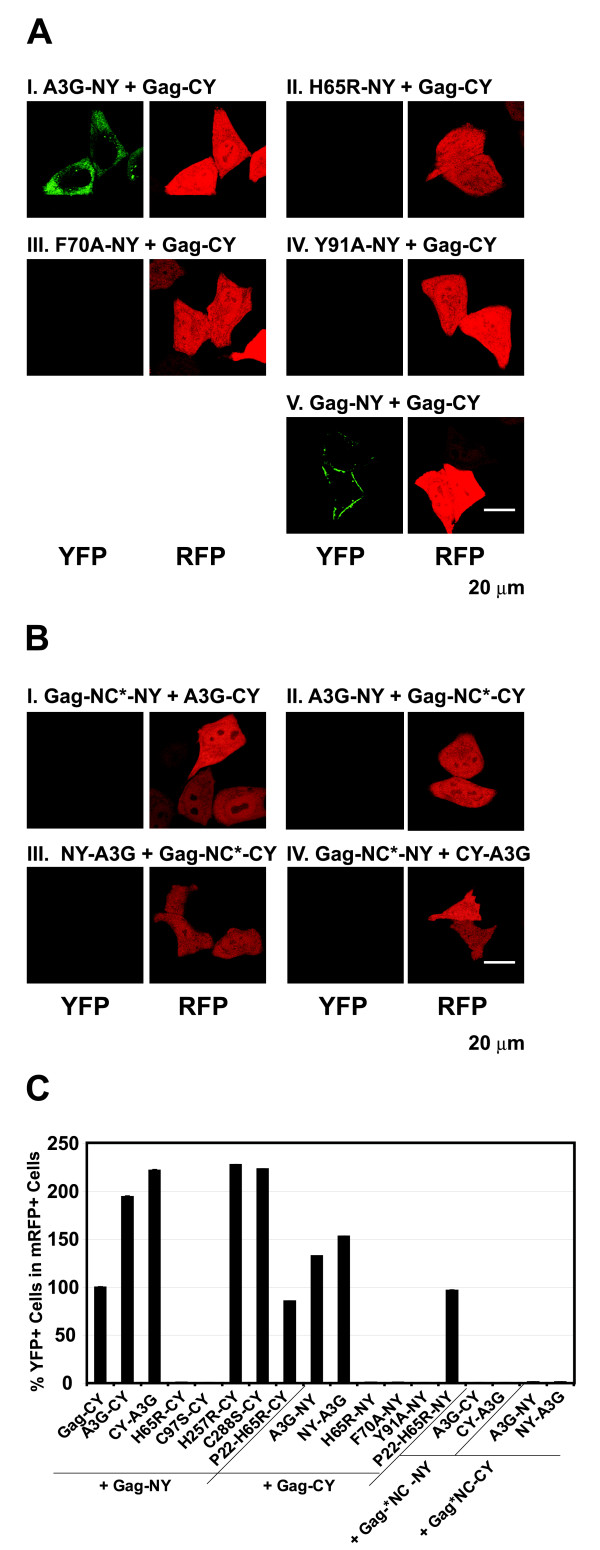
Background: Host restriction factor APOBEC3G (A3G) blocks human immunodeficiency virus type 1 (HIV-1) replication by G-to-A hypermutation, and by inhibiting DNA synthesis and provirus formation. Previous reports have suggested that A3G is a dimer and its virion incorporation is mediated through interactions with viral or nonviral RNAs and/or HIV-1 Gag. We have now employed a bimolecular fluorescence complementation assay (BiFC) to analyze the intracellular A3G-A3G, A3G-RNA, and A3G-Gag interactions in living cells by reconstitution of yellow fluorescent protein (YFP) from its N- or C-terminal fragments.

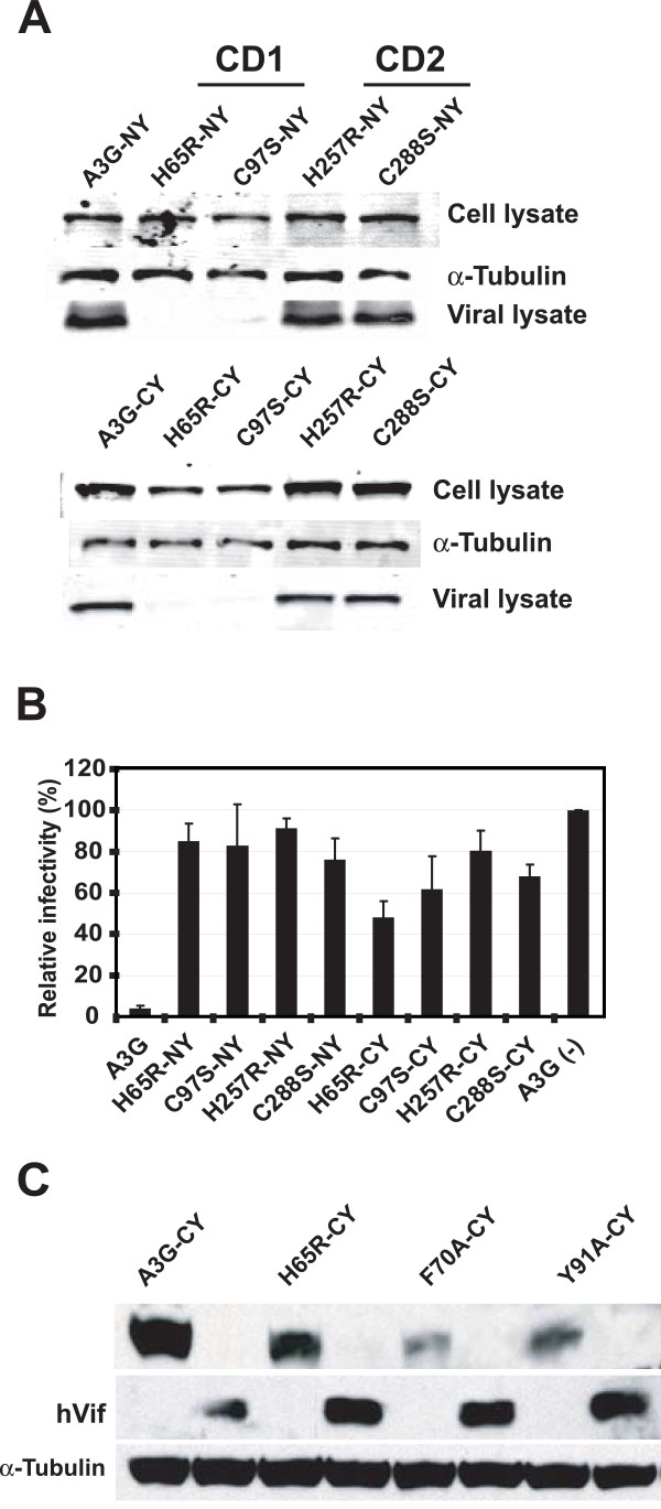
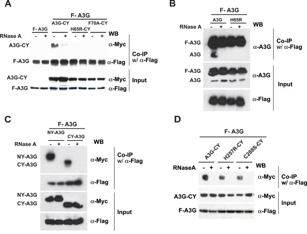
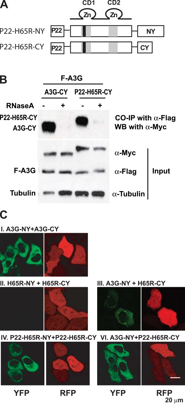
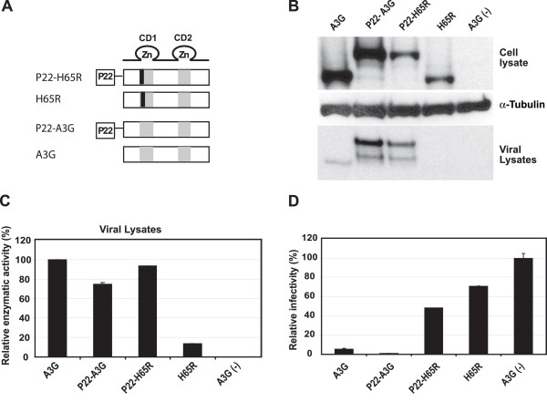
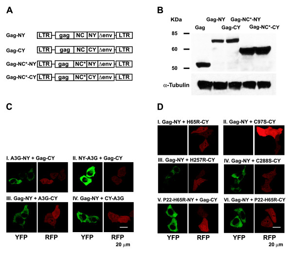
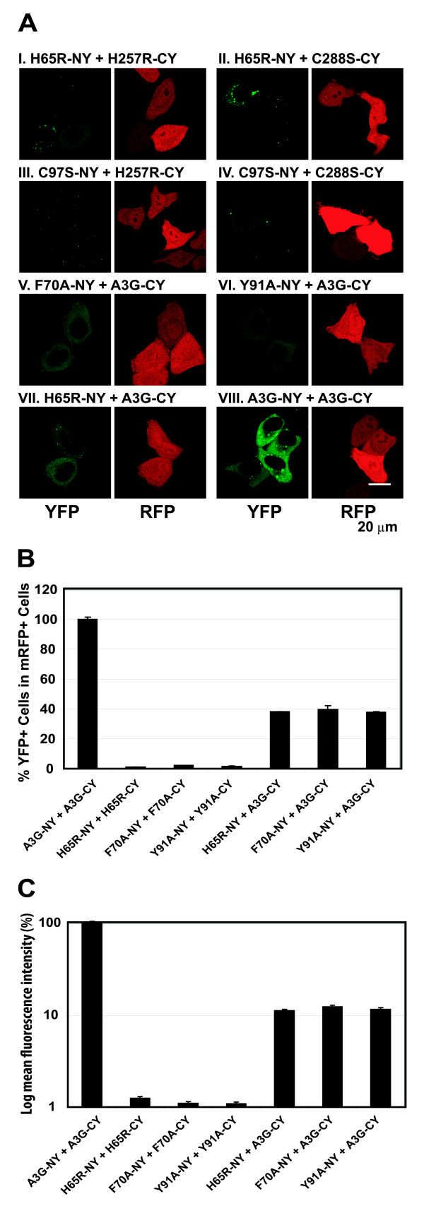
Results: The results obtained with catalytic domain 1 and 2 (CD1 and CD2) mutants indicate that A3G-A3G and A3G-Gag multimerization is dependent on an intact CD1 domain, which is required for RNA binding. A mutant HIV-1 Gag that exhibits reduced RNA binding also failed to reconstitute BiFC with wild-type A3G, indicating a requirement for both HIV-1 Gag and A3G to bind to RNA for their multimerization. Addition of a non-specific RNA binding peptide (P22) to the N-terminus of a CD1 mutant of A3G restored BiFC and virion incorporation, but failed to inhibit viral replication, indicating that the mutations in CD1 resulted in additional defects that interfere with A3G's antiviral activity.

Conclusion: These studies establish a robust BiFC assay for analysis of intracellular interactions of A3G with other macromolecules. The results indicate that in vivo A3G is a monomer that forms multimers upon binding to RNA. In addition, we observed weak interactions between wild-type A3G molecules and RNA binding-defective mutants of A3G, which could explain previously described protein-protein interactions between purified A3G molecules.


Figures

Figure 1
Figure 1
A3G BiFC constructs and their biological activities. (A) Structures of A3G BiFC constructs A3G-NY, A3G-CY, NY-A3G, and CY-A3G. The YFP N-terminal (NY) and C-terminal (CY) fragments were fused either to the C-terminal end of A3G (A3G-NY and A3G-CY) or the N-terminal end of A3G (NY-A3G and CY-A3G). The glycine-rich hinge regions (thin lines) for N-terminally tagged BiFC constructs is slightly longer than in the C-terminally tagged constructs. The catalytic domains 1 and 2 (CD1 and CD2) are shown as gray boxes. (B) Western blotting analysis of cells co-transfected with HDV-EGFP along with wild-type A3G or A3G BiFC constructs. The A3G protein was detected using a polyclonal anti-A3G antibody. (C) Relative cytidine deaminase activity in lysates of cells co-transfected with wild-type A3G or A3G BiFC constructs as well as pHDV-EGFP, pC-HelpΔVif and pHCMV-G. Total cellular protein (0.3 μg) from each cell lysate was used for determination of enzymatic activity, and the activity in cells transfected with wild-type A3G was set to 100%. Error bars represent the standard error of the mean (s.e.m.) of three independent experiments. (D) Effect of wild-type A3G and A3G BiFC constructs on infectivity of HDV-EGFP. The infectivity of the virions produced in the absence and presence of HIV-1 Vif was determined by flow cytometry analysis of cells infected with the virions. Transfections were also performed in the absence of A3G and Vif, and the proportion of GFP+ cells after infection with HDV-EGFP (23.4% in the absence of Vif, and 28% in the presence of Vif) was set to 100%. Error bars represent the s.e.m. of three independent experiments.
Figure 2
Figure 2
Reconstitution of YFP fluorescence with A3G BiFC constructs. (A) Reconstitution of fluorescence upon cotransfection with A3G BiFC constructs. HeLa cells were cotransfected with A3G BiFC constructs and mRFP expression plasmid to identify transfected cells. Fluorescence was reconstituted upon co-transfection with A3G-NY + A3G-CY (I), NY-A3G + CY-A3G (II), A3G-NY + CY-A3G (III), and NY-A3G + A3G-CY (IV). Transfection with A3G-NY (V), A3G-CY (VI), NY-A3G (VII), or CY-A3G (VIII) did not produce YFP fluorescence. (B) Quantfication of BiFC using flow cytometry analysis. 293T cells were co-transfected with A3G BiFC constructs and mRFP expression plasmid as an internal control for transfection, and the percentage of YFP+ cells in mRFP+ cells was determined. The percentage of YFP+ cells in mRFP+ cells after co-transfection with A3G-NY and A3G-CY (15.3%) was set to 100%. The error bars represent the s.e.m. of two independent experiments.
Figure 3
Figure 3
A3G BiFC constructs containing mutations in CD1 or CD2 and their biological activities. (A) Western blotting analysis of lysates of 293T cells and viral lysates produced from cells co-transfected with pHDV-EGFP, pC-HelpΔVif and pHCMV-G and wild-type A3G or A3G BiFC constructs containing mutations in the CD1 (H65R-NY, H65R-CY, C97S-NY, and C97S-CY) or CD2 (H257R-NY, H257R-CY, C288S-NY, and C288S-CY). The cell lysates were also analyzed using anti-tubulin antibody to insure equivalent loading of cell lysate proteins (panel labeled α-tubulin). (B) Effects of CD1 or CD2 mutations on A3G's ability to inhibit HIV-1 replication. 293T cells were co-transfected with wild-type A3G or A3G BiFC constructs along with pHDV-EGFP, pC-HelpΔVif, and pHCMV-G, and the infectivity of the virions produced was determined by flow cytometry analysis of the infected cells for EGFP expression. The proportion of GFP+ cells in the absence of A3G co-transfection was set to 100%. Error bars represent the s.e.m. of three independent experiments. (C) Vif sensitivity of CD1 domain mutants. A3G-CY and CD1 domain mutants H65R-CY, F70A-CY, and Y91A-CY were transfected into 293T cells with and without Vif expression plasmid. A3G fusion proteins were detected by using anti-A3G antibody and HIV-1 Vif was detected using anti-Vif polyclonal antiserum. Anti-tubulin antibody was used to detect tubulin, which served as a loading control.
Figure 4
Figure 4
RNA binding activities of CD1 and CD2 domain mutants of A3G. (A) Effect of CD1 mutations on the ability of A3G to bind cellular RNA. 293T cells were co-transfected with pF-A3G and empty vector, or pF-A3G and A3G-CY, or pF-A3G and fourfold higher amounts of H65R-CY or F70A-CY DNA compared to pF-A3G. An anti-FLAG antibody was used to co-immunoprecipitate F-A3G and associated proteins in the presence or absence of RNase A treatment. The F-A3G, A3G-CY, H65R-CY, and F70A-CY proteins were detected by Western blot using an anti-A3G antibody. (B) A3G-A3G interactions between F-A3G and untagged A3G proteins. 293T cells were co-transfected with F-A3G and empty vector, F-A3G and fourfold higher amounts of untagged A3G DNA or F-A3G and fourfold higher amounts of untagged H65R mutant of A3G DNA. Co-IP assays were performed as described in Fig. 4A. (C) Effect of N-terminal NY and CY tags on A3G-A3G interactions. 293T cells were co-transfected with F-A3G and NY-A3G or CY-A3G. Co-IP assays were performed as described in Fig. 4A. (D) Effect of CD2 mutations on ability of A3G to bind to RNA. 293T cells were co-transfected with pF-A3G and empty vector, or pF-A3G and A3G-CY, or pF-A3G and fourfold higher amounts of H257R-CY, and C288S-CY DNA compared to pF-A3G. Co-IP assays were performed as described in Fig. 4A.
Figure 5
Figure 5
BiFC assays with CD1 and CD2 mutants of A3G. (A) BiFC assays with CD2 mutants of A3G. All co-transfections included mRFP expressing plasmid, and RFP expression was used to identify transfected cells (panels labeled RFP). (B) BiFC and immunofluorescence assays with CD1 mutants of A3G. Expression of the CD1 mutants was verified by detection of the H65R-NY, H65R-CY, C97S-NY, and C97S-CY proteins in transfected cells by immunofluorescence. An anti-A3G polyclonal antibody produced in rabbit was used as a primary antibody and Alexa Fluor 568-conjugated goat antibody to rabbit IgG (H+L) (Molecular Probes) was used as secondary fluorescent antibody. (C) Comparison of BiFC and protein expression between WT A3G and H65R mutant A3G. Western blotting analysis of lysates of cells co-transfected with A3G-NY and -CY (I, 0.25 μg DNA each) or H65R-NY and -CY (II, 1 μg DNA each). The A3G proteins were identified by using a polyclonal anti-A3G antibody, and the same lysates were analyzed by using an anti-tubulin antibody to ensure that equivalent amounts were loaded onto gels. (D) Western blotting analysis of lysates of 293T cells and viral lysates produced from cells transfected with CD1 mutants F70A-NY, F70A-CY, Y91A-NY, and Y91A-CY. The cell lysates were also analyzed using anti-tubulin antibody to insure equivalent loading of cell lysate proteins (panel labeled α-tubulin). (E) BiFC assays with CD1 mutants F70A and Y91A.
Figure 6
Figure 6
Effect of non-specific RNA-binding peptide on BiFC with CD1 mutant H65R. (A) Structure of P22-H65R BiFC constructs. P22 is a 20-amino-acid basic peptide derived from bacteriophage P22 that was fused to the N-terminus of H65R-NY and H65R-CY with a flexible hinge region between P22 and A3G. (B) Effect of P22 peptide on ability of H65R mutant to bind to RNA. 293T cells were co-transfected with pF-A3G and empty vector, or pF-A3G and A3G-CY, or pF-A3G and fourfold higher amount of P22-H65R-CY compared to pF-A3G. Co-IP assays were performed as described for Fig. 4A. (C) BiFC assays to evaluate interactions between wild-type and mutant A3Gs.
Figure 7
Figure 7
Effect of non-specific RNA-binding peptide on encapsidation and antiviral activity of CD1 mutant H65R. (A) Strucures of constructs P22-H65R, H65R, P22-A3G, and wild-type A3G. (B) Western blotting analysis of lysates of 293T cells co-transfected with the A3G constructs as well as pHDV-EGFP, C-HelpΔVif and pHCMV-G (panel labeled Cell lysate). The cell lysates were also analyzed using anti-tubulin antibody to insure equivalent loading of cell lysate proteins (panel labeled α-tubulin). Incorporation of A3G BiFC constructs into VLPs (panel labeled Viral lysate) was determined by analyzing viral lysates containing 100 ng of p24 CA. (C) Relative cytidine deaminase activity in viral lysates of virions produced in the presence of wild type A3G, P22-A3G, P22-H65R, H65R, or in the absence of A3G. The cytidine deaminase activity of A3G-CY was set to 100%. (D) Relative HDV-EGFP infectivity in the presence of P22-A3G, P22-H65R and H65R constructs. Infectivity of the virions produced was determined by flow cytometry analysis of cells infected with the virions. The proportion of GFP+ cells after infection with HDV-EGFP in the absence of A3G was set to 100%. Error bars represent the s.e.m. of three independent experiments.
Figure 8
Figure 8
Characterization of HIV-1 Gag BiFC constructs and interactions with A3G. (A) Structure of HIV-1 Gag BiFC constructs. NC*, RNA-binding defective mutant of HIV-1 Gag. (B) Western blot analysis of HIV-1 Gag expression from BiFC constructs (anti-HIV-Gag polyclonal antibody) and α-tubulin in transfected 293T cells. (C) BiFC assays to evaluate interactions between HIV-1 Gag and wild-type A3G. (D) BiFC assays to evaluate interactions between HIV-1 Gag and CD1 or CD2 domain mutants of A3G.
Figure 9
Figure 9
BiFC assays to evaluate interactions between HIV-1 Gag, A3G, and RNA-binding defective mutants of Gag and A3G. (E) BiFC assays to evaluate interactions between HIV-1 Gag and RNA-binding defective A3G mutants H65R, F70A, and Y91A. (F) BiFC assays to evaluate interactions between HIV-1 Gag NC mutants and A3G. (G) Quantification of YFP fluorescence reconstitution with HIV-1 Gag and A3G BiFC constructs using flow cytometry analysis. The percentage of YFP+ cells in mRFP+ cells after co-transfection with Gag-NY and Gag-CY (3.5%) was set to 100%. The error bars represent the s.e.m. of two independent experiments.
Figure 10
Figure 10
BiFC assays to evaluate interactions between RNA-binding competent A3G proteins and RNA-binding defective A3G proteins. (A) BiFC assays to evaluate interactions between mutant A3Gs. (B) Quantification of BiFC efficiency was determined by flow cytometry analysis as described in Fig. 2B. The percentage of YFP+ cells among the mRFP+ cells after cotransfection with A3G-NY and A3G-CY (15.3%) was set to 100%. The error bars represent the s.e.m. of two independent experiments. (C) Quantification of BiFC fluorescence mean intensity using flow cytometry analysis. The mean intensities of YFP+ cells were determined using CELLQUEST software. The error bars represent the s.e.m. of two independent experiments.

Similar articles

Cited by

References

    1. Sheehy AM, Gaddis NC, Choi JD, Malim MH. Isolation of a human gene that inhibits HIV-1 infection and is suppressed by the viral Vif protein. Nature. 2002;418:646–650. - PubMed
    1. Stremlau M, Owens CM, Perron MJ, Kiessling M, Autissier P, Sodroski J. The cytoplasmic body component TRIM5alpha restricts HIV-1 infection in Old World monkeys. Nature. 2004;427:848–853. - PubMed
    1. Harris RS, Bishop KN, Sheehy AM, Craig HM, Petersen-Mahrt SK, Watt IN, Neuberger MS, Malim MH. DNA deamination mediates innate immunity to retroviral infection. Cell. 2003;113:803–809. - PubMed
    1. Mangeat B, Turelli P, Caron G, Friedli M, Perrin L, Trono D. Broad antiretroviral defence by human APOBEC3G through lethal editing of nascent reverse transcripts. Nature. 2003;424:99–103. - PubMed
    1. Lecossier D, Bouchonnet F, Clavel F, Hance AJ. Hypermutation of HIV-1 DNA in the absence of the Vif protein. Science. 2003;300:1112. - PubMed

Publication types