. 2009 Jun 4:9:120.
doi: 10.1186/1471-2180-9-120.

Expression and subcellular localization of kinetoplast-associated proteins in the different developmental stages of Trypanosoma cruzi

Affiliations

Expression and subcellular localization of kinetoplast-associated proteins in the different developmental stages of Trypanosoma cruzi

Danielle Pereira Cavalcanti et al. BMC Microbiol. .

Abstract

Background: The kinetoplast DNA (kDNA) of trypanosomatids consists of an unusual arrangement of circular molecules catenated into a single network. The diameter of the isolated kDNA network is similar to that of the entire cell. However, within the kinetoplast matrix, the kDNA is highly condensed. Studies in Crithidia fasciculata showed that kinetoplast-associated proteins (KAPs) are capable of condensing the kDNA network. However, little is known about the KAPs of Trypanosoma cruzi, a parasitic protozoon that shows distinct patterns of kDNA condensation during their complex morphogenetic development. In epimastigotes and amastigotes (replicating forms) the kDNA fibers are tightly packed into a disk-shaped kinetoplast, whereas trypomastigotes (non-replicating) present a more relaxed kDNA organization contained within a rounded structure. It is still unclear how the compact kinetoplast disk of epimastigotes is converted into a globular structure in the infective trypomastigotes.

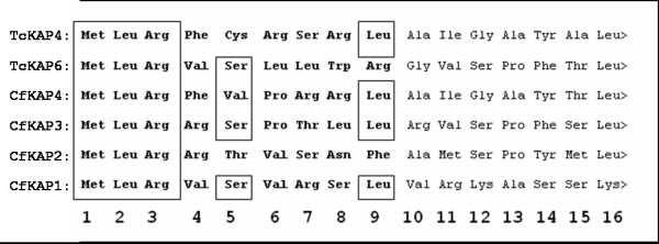
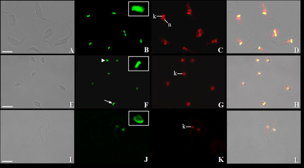
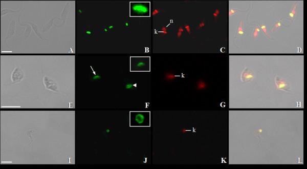
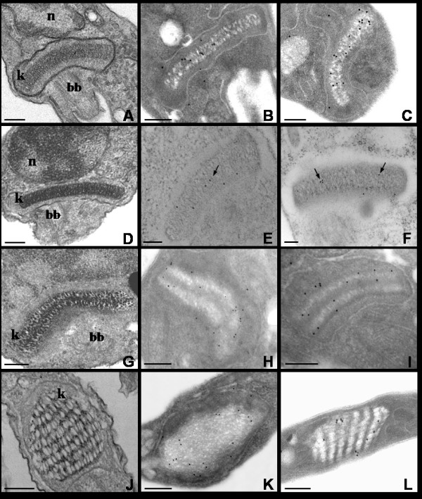
Results: In this work, we have analyzed KAP coding genes in trypanosomatid genomes and cloned and expressed two kinetoplast-associated proteins in T. cruzi: TcKAP4 and TcKAP6. Such small basic proteins are expressed in all developmental stages of the parasite, although present a differential distribution within the kinetoplasts of epimastigote, amastigote and trypomastigote forms.

Conclusion: Several features of TcKAPs, such as their small size, basic nature and similarity with KAPs of C. fasciculata, are consistent with a role in DNA charge neutralization and condensation. Additionally, the differential distribution of KAPs in the kinetoplasts of distinct developmental stages of the parasite, indicate that the kDNA rearrangement that takes place during the T. cruzi differentiation process is accompanied by TcKAPs redistribution.


Figures

Figure 1
Figure 1
Phylogenetic analysis of trypanosomatid KAPs proteins with confidence values shown as percentages. Lb, Leishmania braziliensis; Li, Leishmania infantum; Lm, Leishmania major; Tb, Trypanosoma brucei; Tc, Trypanosoma cruzi.
Figure 2
Figure 2
Comparison of N-terminal sequences of KAPs from C. fasciculata and T. cruzi. The presequences predicted to be involved in kinetoplast import are shown in bold type. The boxes indicate the highly conserved amino acids. Note that all sequences begin with the sequence M, L, R. In all sequences other than those of CfKAP4 and TcKAP4, the fifth amino acid is hydroxylated and the ninth is generally hydrophobic. CfKAP4 (PIR JC6092), CfKAP3 (GenBank accession number AY143553), CfKAP2 (GenBank accession numbers AF008943 and AF008944) and CfKAP1 (GenBank accession number AF034951) are KAPs from C. fasciculata whereas TcKAP4 (GenBank accession number ABR15473) and TcKAP6 (GenBank accession number ABR15474) are T. cruzi KAPs.
Figure 3
Figure 3
Expression of recombinant TcKAPs in E. coli. The TcKAP4 (A) and TcKAP6 (B) were expressed in E. coli M15 strain following induction with 1 mM IPTG for 3 h. Immunoblotting assays of non-induced (1) and induced (2) bacterial extract using anti-polyhistidine antibody confirmed the expression of recombinant TcKAPs.
Figure 4
Figure 4
Detection of TcKAPs in T. cruzi. Western blot analyses of (1) epimastigote, (2) amastigote/intermediate form and (3) trypomastigote extracts of T. cruzi, using anti-TcKAP4 (A) or anti-TcKAP6 (B) serum. Both antisera recognized a single polypeptide in all developmental stages of the parasite.
Figure 5
Figure 5
Distribution of TcKAP4 in T. cruzi. Immunolocalization of TcKAP4 in epimastigotes (A-D), amastigotes/intermediate forms (E-H) and trypomastigotes (I-L) of T. cruzi. In epimastigotes (B) and amastigotes (F-arrow), the protein is distributed throughout the kDNA disk (insets). In intermediate forms (F-arrowhead) and trypomastigotes (J-inset), a peripheral labeling of the kinetoplast was observed. (A-E-I) Phase-contrast image, (B-F-J) fluorescence image using anti-TcKAP4 serum, (C-G-K) propidium iodide showing the nucleus (n) and the kinetoplast (k), and (D-H-L) the overlay image. Bars = 5 μm.
Figure 6
Figure 6
Distribution of TcKAP6 in T. cruzi. Immunolocalization of TcKAP6 in epimastigotes (A-D), amastigotes/intermediates forms (E-H) and trypomastigotes (I-L) of T. cruzi. As observed for TcKAP4, this protein was also distributed throughout kDNA disk in epimastigotes (B-inset) and amastigotes (F-arrow and inset), and at the periphery of the kinetoplast in intermediate forms (F-arrowhead) and trypomastigotes (J-inset). (A-E-I) Phase-contrast image, (B-F-J) location of TcKAP6 in the kinetoplasts of T. cruzi, (C-G-K) iodide propidium labeling and (D-H-L) the overlay image. k = kinetoplast, n = nucleus. Bars = 5 μm.
Figure 7
Figure 7
Ultrastructural localization of TcKAPs by transmission electron microscopy. A-D-G-J: ultrastructural analyses of the kinetoplast in the different developmental stages of T. cruzi. The kinetoplast of intermediate forms (G) is larger than the bar-shaped kinetoplast of epimastigotes (A) and amastigotes (D). The trypomastigotes (J) present a more relaxed kDNA organization, contained within a rounded kinetoplast. TcKAP4 (B-E-H-K) was distributed throughout the kinetoplast DNA network in epimatigotes (B) and amastigotes (E-arrow). In intermediate forms (H) and in trypomastigotes (K), TcKAP4 was distributed mainly at the periphery of the kDNA. The same result was observed for TcKAP6 (C-F-I-L). A homogenous distribution for all kinetoplast was observed in epimastigotes (C) and amastigotes (F-arrows), while a more peripherical distribution was seen in intermediate forms (I) and trypomastigotes (L). Bars = 0.25 μm. k = kinetoplast, n = nucleus, bb = basal body.

References

    1. Kornberg RD, Lorch Y. Twenty-five years of the nucleosome, fundamental particle of the eukaryote chromosome. Cell. 1999;98:285–294. - PubMed
    1. Polo SE, Almouzni G. Chromatin assembly: a basic recipe with various flavours. Curr Opin Genet Dev. 2006;16:104–111. - PubMed
    1. Sandman K, Reeve JN. Archaeal chromatin proteins: different structures but common function? Curr Opin Microbiol. 2005;8:656–661. - PubMed
    1. Luijsterburg MS, Noom MC, Wuite GJ, Dame RT. The architectural role of nucleoid-associated proteins in the organization of bacterial chromatin: a molecular perspective. J Struct Biol. 2006;156:262–272. - PubMed
    1. Shapiro TA, Englund PT. The structure and replication of kinetoplast DNA. Annu Rev Microbiol. 1995;49:117–43. - PubMed

Publication types

LinkOut - more resources