. 2009 Aug 15;332(2):212-22.
doi: 10.1016/j.ydbio.2009.05.570. Epub 2009 Jun 2.

Dynamic positional fate map of the primary heart-forming region

Affiliations

Dynamic positional fate map of the primary heart-forming region

Cheng Cui et al. Dev Biol. .

Abstract

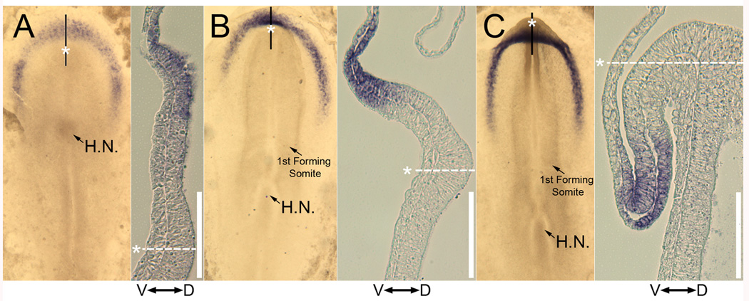
Here we show the temporal-spatial orchestration of early heart morphogenesis at cellular level resolution, in vivo, and reconcile conflicting positional fate mapping data regarding the primary heart-forming field(s). We determined the positional fates of precardiac cells using a precision electroporation approach in combination with wide-field time-lapse microscopy in the quail embryo, a warm-blooded vertebrate (HH Stages 4 through 10). Contrary to previous studies, the results demonstrate the existence of a "continuous" circle-shaped heart field that spans the midline, appearing at HH Stage 4, which then expands to form a wide arc of progenitors at HH Stages 5-7. Our time-resolved image data show that a subset of these cardiac progenitor cells do not overlap with the expression of common cardiogenic factors, Nkx-2.5 and Bmp-2, until HH Stage 10, when a tubular heart has formed, calling into question when cardiac fate is specified and by which key factors. Sub-groups and anatomical bands (cohorts) of heart precursor cells dramatically change their relative positions in a process largely driven by endodermal folding and other large-scale tissue deformations. Thus, our novel dynamic positional fate maps resolve the origin of cardiac progenitor cells in amniotes. The data also establish the concept that tissue motion contributes significantly to cellular position fate - i.e., much of the cellular displacement that occurs during assembly of a midline heart tube (HH Stage 9) is NOT due to "migration" (autonomous motility), a commonly held belief. Computational analysis of our time-resolved data lays the foundation for more precise analyses of how cardiac gene regulatory networks correlate with early heart tissue morphogenesis in birds and mammals.


Figures

Figure 1
Figure 1
Colors were assigned to tracked heart precursor cells in quail embryos to denote their positions at HH stage 10 along both the antero-posterior (A-P) and the ventral-dorsal (V-D) axes. Fluorescent tracked precardiac cells were selected from a population of electroporated epiblastic cells that gastrulated, formed splanchnic mesodermal cells, and came to reside on the surface of the tubular heart. A) A fluorescent image of an HH stage 10 heart with tracked cells labeled with grayscale (intensity) values. To simplify, we assume the heart tube is a cylinder with a radius R: defined as the distance from the midline reference points (arrow-heads) to lateral reference points (arrows) at successive A-P levels; and that all tracked cells are on the cylinder’s surface. The radius R represents the longest distance between a lateral point (white arrow) and the corresponding midline point (white arrowhead) at the same A-P level. The radius measurement can vary at different A-P levels. B) The distance (x) between a tracked cell and the midline position of the heart tube at the same A-P level is used to calculate the tracked cell’s position on the cylinder surface (c) using the equation shown. C) The tracked cell is then assigned one color according to its position on the cylinder’s surface to indicate its relative position along the V-D axis. The color red represents the ventral-most positions; the color blue represents the dorsal-most positions and the color green represents the lateral-most positions. Along the A-P axis, the tracked cells are assigned a separate color according to their positions relative to two reference points: A, the anterior-most point and B, the posterior-most point of the heart tube (see panel 1A asterisks). The color red represents the anterior-most positions; the color blue represents the posterior-most positions and the color green represents the middle positions. The cells that reside caudal to the heart (future omphalomesenteric veins) by HH stage 10 (cells in the white circle in panel A) are assigned the color pink (See Figure 2 and Movie 3).
Figure 2
Figure 2
Sequential time-lapse frames of “tracked” primary cardiac precursor cells compiled from 6 quail embryos between HH stages 4 through 9. The tracked cells are pseudo-colored and super-imposed upon a corresponding bright-field image (Panels A-F). The color code denotes a cell’s final position in the HH10 heart tube with respect to the A-P axis, as explained in Figure 1. The cranial-most cells were tracked on both sides of the embryonic vertebral axis (see white arrows); while cells originating inside the region depicted by the white oval (Panel A) were tracked only on the embryonic left side. The colored radian lines emanating from Hensen’s node (HN) denote the spatial boundaries of the color-coded cells at each time point. See Movie 3 for the corresponding time-lapse data. The columns of smaller image panels depict a series of whole-mounted embryos subjected to RNA in-situ hybridizations using Bmp-2 or Nkx-2.5 probes. In situ hybridizations are shown in quail embryos for Nkx-2.5, and in chicken embryos for Bmp-2. Cells colored pink will reside caudal to the heart (future omphalomesenteric veins) by HH stage 10 and will eventually contribute to the atrium. Black arrows indicate a subset of pink cells that do not overlap with Nkx-2.5 expression. Scale bar: 200 µm.
Figure 3
Figure 3
Sequential time points show cellular motion analyzed with respect to the dorsoventral, D-V, axis. The panels are arranged similar to Figure 2, in which primary cardiac precursor cells from 6 quail embryos were tracked between HH stage 4 and 9 (A-F). The color code denotes a cell’s final position in the HH10 heart tube with respect to the D-V axis, as explained in Figure 1. The cranial-most cells were tracked on both sides of the embryonic axis (as indicated in Figure 2), while only left side cells were tracked for the D-V analysis (Movie 3). The colored radian lines emanating from Hensen’s node (HN) denote the spatial boundaries of the color-coded cells at each time point. RNA in situ hybridization panels are shown to the right of each respective time-lapse frame. The corresponding in situ hybridizations for Nkx2.5 are shown in chicken embryos. Bmp-2 images are duplicates of those seen in Figure 2.
Figure 4
Figure 4
Anterior-most cardiac precursor cells overlap with Bmp-2 expression patterns at HH5 and HH7. The image in panel A depicts an HH4- embryo showing only cells fated to assume cranial-most heart positions at subsequent stages (See Movie 4 and Movie 5). Panel B and C are duplicates of Figure 2 HH5 and HH7, with the black boxes showing a region expressing Bmp-2. Panels B' and C' show the boxed regions at higher magnification.
Figure 5
Figure 5
Images of whole-mount embryos (WM-BF and WM-GFP) and their longitudinal-sections (DIC and GFP) at HH5 (A), HH6 (B) and HH7+ (C). Quail embryos were electroporated with mito-YFP at HH 3 and whole-mount (WM) immunolabeled with anti-GFP antibody to enhance the fluorescence signal before sectioning. The longitudinal-sections are marked as solid white lines in WM-GFP images. The A-P levels of cardiac precursor cells that reside at the cranial-most positions, as suggested in our time-lapse data, are marked with black asterisks in WM-BF images and with white dashed lines and asterisks in images of longitudinal-sections. White arrowheads mark the mesodermal cells. The embryos in whole-mount images are viewed from their ventral side. Double-headed arrows with letters ‘V’ and ‘D’ in DIC images of longitudinal-sections indicate the ventral and dorsal directions of the sections. The white boxes indicate the corresponding regions of the forming headfold at each time shown. The boxed region in Panel C is referred to in the text as the lip of the headfold. White scale bars: 100 µm. HN = Hensen’s node; PS = primitive streak
Figure 6
Figure 6
Whole-mount embryo RNA in-situ hybridization for Nkx-2.5 and longitudinal-section images of avian embryos at HH5+ (A; chick), HH6+ (B; quail) and HH7+ (C; quail). The black lines in whole-mounted (WM) in-situ images indicate the site of longitudinal-sections. White dashed lines and asterisks indicate the A-P levels of cardiac precursor cells that reside at the anterior-most positions as suggested in our time-lapse data. The whole-mounted embryos are viewed from their ventral side. Double headed arrows with letters ‘V’ and ‘D’ in longitudinal-section images indicate the ventral and dorsal directions of the sections. White scale bars: 100 µm.
Figure 7
Figure 7
Schematic representation of the spatial separation of pseudo-colored (based on cell position at HH 10) cells at HH7 along the A-P (viewer’s left side; hatched) and the V-D (viewer’s right side) axes of the embryo. The color pink represents the region containing cells that will contribute predominantly to the atria by HH12. These data represent cell tracking in 6 embryos. (See also Movie 1–Movie 4).

References

    1. Abu-Issa R, Kirby ML. Heart Field: From Mesoderm to Heart Tube. Annu Rev Cell Dev Biol. 2007 - PubMed
    1. Abu-Issa R, Kirby ML. Patterning of the heart field in the chick. Dev. Biol. 2008;319:223–233. - PMC - PubMed
    1. Abu-Issa R, Waldo K, Kirby ML. Heart fields: one, two or more? Dev Biol. 2004;272:281–285. - PubMed
    1. Baldwin HS, Jensen KL, Solursh M. Myogenic cytodifferentiation of the precardiac mesoderm in the rat. Differentiation. 1991;47:163–172. - PubMed
    1. Campbell RE, Tour O, Palmer AE, Steinbach PA, Baird GS, Zacharias DA, Tsien RY. A monomeric red fluorescent protein. Proc Natl Acad Sci U S A. 2002;99:7877–7882. - PMC - PubMed

Publication types

MeSH terms

LinkOut - more resources