Review
. 2009 Jul;27(7):423-33.
doi: 10.1016/j.tibtech.2009.04.002. Epub 2009 Jun 6.

Production of self-assembling biomaterials for tissue engineering

Affiliations
Review

Production of self-assembling biomaterials for tissue engineering

Stuart Kyle et al. Trends Biotechnol. 2009 Jul.

Abstract

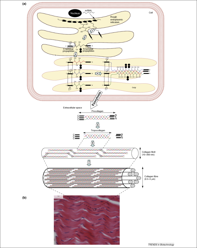
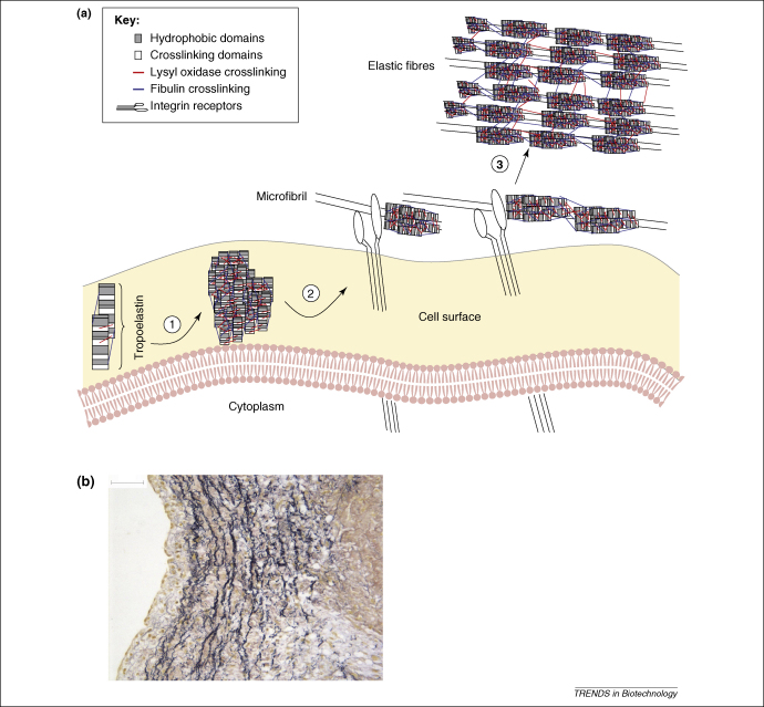
Self-assembling peptide-based biomaterials are being developed for use as 3D tissue engineering scaffolds and for therapeutic drug-release applications. Chemical synthesis provides custom-made peptides in small quantities, but production approaches based upon transgenic organisms might be more cost-effective for large-scale peptide production. Long lead times for developing appropriate animal clones or plant lines and potential negative public opinion are obstacles to these routes. Microbes, particularly safe organisms used in the food industry, offer a more rapid route to the large-scale production of recombinant self-assembling biomaterials. In this review, recent advances and challenges in the recombinant production of collagen, elastin and de novo designed self-assembling peptides are discussed.


Figures

Figure 1
Figure 1
Collagen biosynthesis. (a) This panel shows the major events in collagen fibre assembly. Modifications of collagen include hydroxylation of prolyl and lysyl residues (1), addition of N-linked oligosaccharides (1) and glycosylation of hydroxylysyl residues in the endoplasmic reticulum (2), before chain alignment and disulphide bond formation (3), which results in the formation of the procollagen triple helix in the Golgi (4). After export from the cell, the N- and C-terminal propeptides are cleaved (5) and the resulting tropocollagen undergoes extensive crosslinking and self-assembly into collagen fibrils of diameters between 10 and 300 nm (6). These fibres in turn further assemble into larger fibres (0.5–3 μm diameter) (7). (b) Histological section of porcine patellar tendon showing a large number of intertwined collagen fibres with ligament fibroblasts, which are stained with haemotoxylin (stains nuclei in blue-purple) and eosin (stains collagen fibres in pink). The scale bar represents 25 μm.
Figure 2
Figure 2
Elastin fibres. (a) This panel schematically illustrates the formation of elastic fibres and their assembly. After translation and prolyl hydroxylation, tropoelastin is transported to the cell surface membrane, where crosslinking by lysyl oxidase aids in tropoelastin aggregation (1). Cell surface proteins might assist the initial assembly with microfibrillar proteins, such as fibulin-4 and/or fibulin-5, thereby facilitating crosslinking. The resulting aggregates remain on the cell surface before they are transferred to extracellular microfibrils, which interact with the cell through integrins (2). Several microfibrillar proteins interact with tropoelastin, which might help in the transfer of aggregates to the microfibril. Elastin aggregates assembled on the microfibril coalesce into larger structures under possible participation of fibulin-4 and/or fibulin-5. Additional crosslinking by lysyl oxidase then leads to completion of the elastic fibre (3). Adapted from with permission. (b) Histological section of a porcine aortic valve leaflet stained with Miller's elastin stain. The elastic fibre entanglements are shown in dark blue/black. The scale bar represents 50 μm.
Figure 3
Figure 3
Schematic illustration of various self-assembling peptide systems . Molecular ‘Lego’ forms β-sheet structures through interactions between hydrophobic and hydrophilic domains. These peptide systems are able to undergo complementary ionic bonding with the hydrophilic surface. Molecular ‘switches’ change their molecular structure upon changes in temperature. Molecular ‘Velcro’ forms monolayers on surfaces through covalent bonds between a cysteine anchor and gold atoms on the surface. Surfactant-like peptides can undergo self-assembly to form either nanovesicles or nanotubes, as shown.
Figure 4
Figure 4
Example of the tandem-repeat strategy for high yield peptide production in the pET31b expression vector. A fusion protein comprising ketosteroid isomerase (KSI) is targeted to inclusion bodies in E. coli together with tandem repeat peptides encoding the P11-4 sequence and a His tag. The fusion protein and peptides are cleaved with cyanogen bromide (CNBr), which acts on intervening methionine residues. This results in the presence of a C-terminal homoserine lactone (hsl) on each resulting peptide monomer.

Similar articles

Cited by

References

    1. Zhang S. Emerging biological materials through molecular self-assembly. Biotechnol. Adv. 2002;20:321–339. - PubMed
    1. Mitraki A., van Raaij M.J. Folding of β-structured fibrous proteins and self-assembling peptides. Methods Mol. Biol. 2005;300:125–140. - PubMed
    1. Kluge J.A. Spider silks and their applications. Trends Biotechnol. 2008;26:244–251. - PubMed
    1. Köster S. An in situ study of collagen self-assembly processes. Biomacromolecules. 2008;9:199–207. - PMC - PubMed
    1. Shapiro F. Bone development and its relation to fracture repair. The role of mesenchymal osteoblasts and surface osteoblasts. Eur. Cell. Mater. 2008;15:53–76. - PubMed

Publication types