

doi: 10.1109/TBME.2009.2024077.
Epub 2009 Jun 2.
A surface topology and motion compensation system for microsurgery guidance and intervention based on common-path optical coherence tomography
Affiliations
- PMID: 19497807
- PMCID: PMC2846755
- DOI: 10.1109/TBME.2009.2024077
Item in Clipboard
A surface topology and motion compensation system for microsurgery guidance and intervention based on common-path optical coherence tomography
IEEE Trans Biomed Eng.
2009 Sep.
Abstract
A surface topology and motion compensation system for microsurgery guidance and intervention is developed based on common-path optical coherence tomography. A 1-D erosion-based edge-searching method and autoregressive predictor are applied to A-scan data for real-time depth tracking. Images using the topology and motion compensation technique are obtained. In addition, the motion compensation properties are studied. The system can be easily integrated with microsurgery tools and can be used for various clinical applications.
Figures
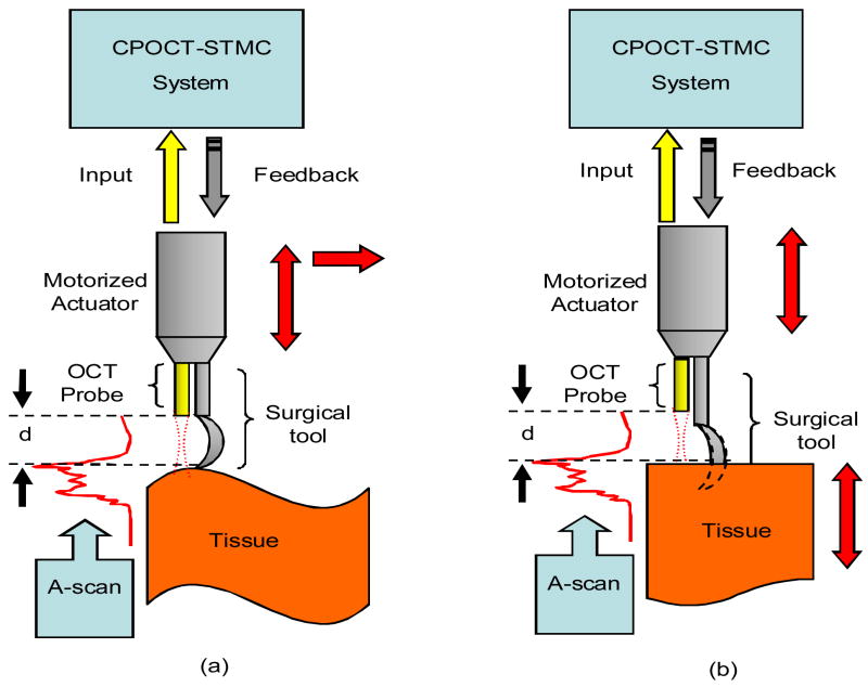
Schematic of CPOCT-STMC system. (a) Topological compensation. (b) Motion compensation.

Edge-searching method. (a) Raw A-scan data, “Ghost edge” exists. (b) A-scan data after 1-D erosion algorithm, “Ghost Edge” is erased and real edge remains. (c) Thresholding of A-scan. (d) First zero-crossing for edge location (depth scale changed).

CPOCT-STMC system flowchart. ASA: A-scan acquisition; SR: surface recognition; AR: autoregressive predictive filter; PC: probe control.

CPOCT-STMC experimental setup.

Images of a phantom sample by CPOCT-STMC system. (a) Traditional OCT with limited imaging depth. (b) Topological compensation with extended imaging depth.

Motion compensation properties of the CPOCT-SMTC system. (a) Response of CPOCT-STMC to a target motion without predictor. (b) Response of CPOCT-STMC to a target motion with AR predictor. (c) Phase difference between response and motion increases with frequency. (d) Response-to-motion amplitude ratio with frequency changing.

Motion compensation of a 3-layer flat phantom sample by CPOCT-STMC system. (a) Experiment details. M: moving stage with motorized actuator; P: fiber CPOCT probe; (b) Traditional B-scan image of static phantom. (c) Traditional B-scan image of periodically moving phantom without depth tracking. (d) STMC B-scan image of periodically moving phantom without predictor. (e) STMC B-scan image of periodically moving phantom with AR predictor.
References
-
- Bouma BE, Tearney GJ. Handbook of Optical Coherence Tomography. Marcel Dekker Inc; NY: 2001. pp. 613–647.
-
- Jafri MS, Tang R, Tang C-M. Optical coherence tomography guided neurosurgical procedures in small rodents. J Neuroscience Methods. 2009 Jan;176:85–95. - PubMed
-
- Low A, Tearney G, Bouma B, Jang I. Technology insight: optical coherence tomography—current status and future development. Nat Clin Pract Cardiovasc Med. 2006 March;3:154–162. - PubMed
-
- Boppart SA, Brezinski ME, Pitris C, Fujimoto JG. Optical Coherence Tomography for Neurosurgical Imaging of Human Intracortical Melanoma. Neurosurgery. 1998 Oct;43:834–841. - PubMed
MeSH terms
Grants and funding
LinkOut - more resources
Full Text Sources
Other Literature Sources
