

DLX5 (distal-less homeobox 5) promotes tumor cell proliferation by transcriptionally regulating MYC
- PMID: 19497851
- PMCID: PMC2742824
- DOI: 10.1074/jbc.M109.021477
DLX5 (distal-less homeobox 5) promotes tumor cell proliferation by transcriptionally regulating MYC
Abstract
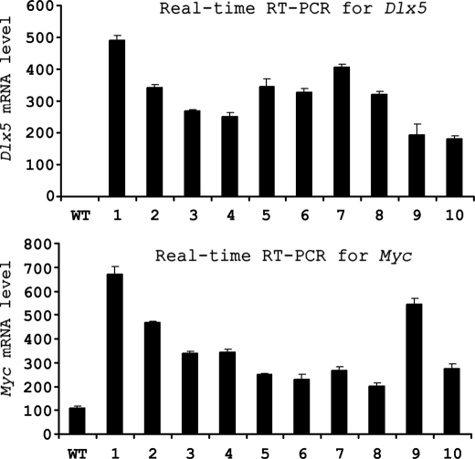
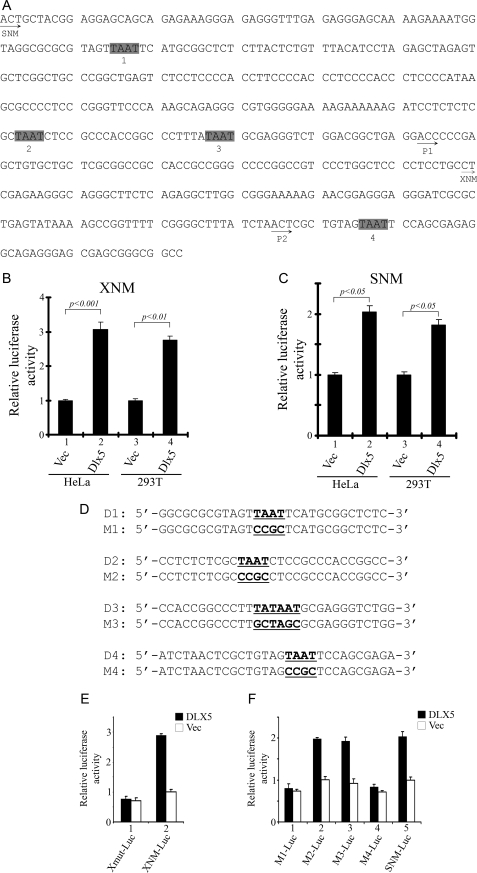
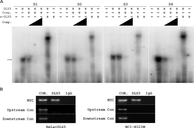
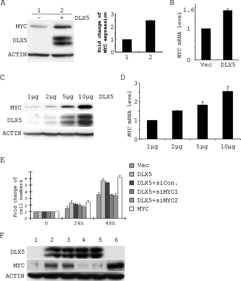
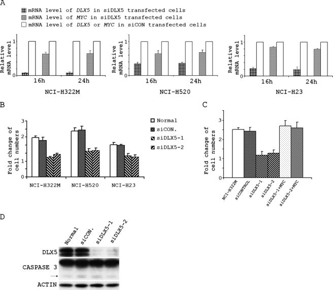
The human DLX homeobox genes, which are related to Dll (Drosophila distal-less gene), encode transcription factors that are expressed primarily in embryonic development. Recently, DLX5 was reported to act as an oncogene in lymphomas and lung cancers, although the mechanism is not known. The identification of target genes of DLX5 can facilitate our understanding of oncogenic mechanisms driven by overexpression of DLX5. The MYC oncogene is aberrantly expressed in many human cancers and regulates transcription of numerous target genes involved in tumorigenesis. Here we demonstrate by luciferase assay that the MYC promoter is specifically activated by overexpression of DLX5 and that two DLX5 binding sites in the MYC promoter are important for transcriptional activation of MYC. We also show that DLX5 binds to the MYC promoter both in vitro and in vivo and that transfection of a DLX5 expression plasmid promotes the expression of MYC in a dose-dependent manner in mammalian cells. Furthermore, overexpression of DLX5 results in increased cell proliferation by up-regulating MYC. Knockdown of DLX5 in lung cancer cells overexpressing DLX5 resulted in decreased expression of MYC and reduced cell proliferation, which was rescued by overexpression of MYC. Because DLX5 has a restricted pattern of expression in adult tissues, it may serve as a potential therapeutic target for the treatment of cancers that overexpress DLX5.
Figures
Similar articles
-
Co-targeting of Akt and Myc inhibits viability of lymphoma cells from Lck-Dlx5 mice.Cancer Biol Ther. 2015;16(4):580-8. doi: 10.1080/15384047.2015.1018495. Epub 2015 Mar 20. Cancer Biol Ther. 2015. PMID: 25793663 Free PMC article.
-
Upregulation of DLX5 promotes ovarian cancer cell proliferation by enhancing IRS-2-AKT signaling.Cancer Res. 2010 Nov 15;70(22):9197-206. doi: 10.1158/0008-5472.CAN-10-1568. Epub 2010 Nov 2. Cancer Res. 2010. PMID: 21045156 Free PMC article.
-
The homeoprotein Dlx5 drives murine T-cell lymphomagenesis by directly transactivating Notch and upregulating Akt signaling.Oncotarget. 2017 Feb 28;8(9):14941-14956. doi: 10.18632/oncotarget.14784. Oncotarget. 2017. PMID: 28122332 Free PMC article.
-
DLX Genes: Roles in Development and Cancer.Cancers (Basel). 2021 Jun 15;13(12):3005. doi: 10.3390/cancers13123005. Cancers (Basel). 2021. PMID: 34203994 Free PMC article. Review.
-
Gene Transactivation and Transrepression in MYC-Driven Cancers.Int J Mol Sci. 2021 Mar 27;22(7):3458. doi: 10.3390/ijms22073458. Int J Mol Sci. 2021. PMID: 33801599 Free PMC article. Review.
Cited by
-
Co-targeting of Akt and Myc inhibits viability of lymphoma cells from Lck-Dlx5 mice.Cancer Biol Ther. 2015;16(4):580-8. doi: 10.1080/15384047.2015.1018495. Epub 2015 Mar 20. Cancer Biol Ther. 2015. PMID: 25793663 Free PMC article.
-
Allelic Switching of DLX5, GRB10, and SVOPL during Colorectal Cancer Tumorigenesis.Int J Genomics. 2019 Apr 10;2019:1287671. doi: 10.1155/2019/1287671. eCollection 2019. Int J Genomics. 2019. PMID: 31093489 Free PMC article.
-
ARF alters PAF1 complex integrity to selectively repress oncogenic transcription programs upon p53 loss.Mol Cell. 2024 Dec 5;84(23):4538-4557.e12. doi: 10.1016/j.molcel.2024.10.020. Epub 2024 Nov 11. Mol Cell. 2024. PMID: 39532099 Free PMC article.
-
Lung Cancer Gene Regulatory Network of Transcription Factors Related to the Hallmarks of Cancer.Curr Issues Mol Biol. 2023 Jan 5;45(1):434-464. doi: 10.3390/cimb45010029. Curr Issues Mol Biol. 2023. PMID: 36661515 Free PMC article.
-
Homeobox Gene Deregulation: Impact on the Hallmarks of Cancer.Cancer Hallm. 2013 Sep 1;1(2-3):67-76. doi: 10.1166/ch.2013.1007. Cancer Hallm. 2013. PMID: 24761365 Free PMC article.
References
-
- Pelengaris S., Khan M. (2003) Arch. Biochem. Biophys. 416, 129–136 - PubMed
-
- Yamada H., Sakamoto H., Taira M., Nishimura S., Shimosato Y., Terada M., Sugimura T. (1986) Jpn. J. Cancer Res. 77, 370–375 - PubMed
-
- Sikora K., Chan S., Evan G., Gabra H., Markham N., Stewart J., Watson J. (1987) Cancer 59, 1289–1295 - PubMed
Publication types
MeSH terms
Substances
Grants and funding
LinkOut - more resources
Full Text Sources
Molecular Biology Databases
Research Materials
