. 2009 Jun 16;106(24):9842-7.
doi: 10.1073/pnas.0904464106. Epub 2009 Jun 3.

Formyl peptide receptors are candidate chemosensory receptors in the vomeronasal organ

Affiliations

Formyl peptide receptors are candidate chemosensory receptors in the vomeronasal organ

Stephen D Liberles et al. Proc Natl Acad Sci U S A. .

Abstract

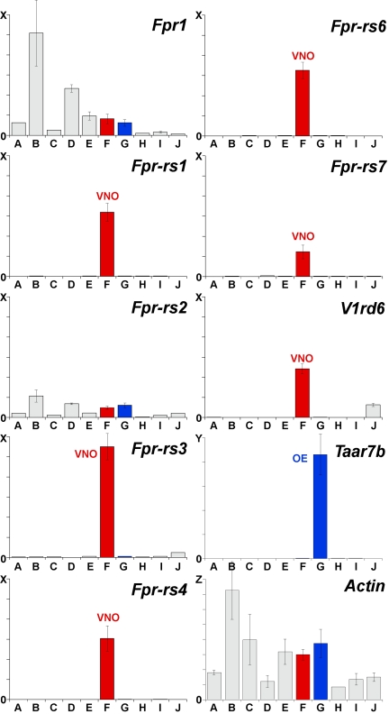
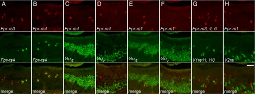
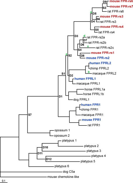
The identification of receptors that detect environmental stimuli lays a foundation for exploring the mechanisms and neural circuits underlying sensation. The mouse vomeronasal organ (VNO), which detects pheromones and other semiochemicals, has 2 known families of chemoreceptors, V1Rs and V2Rs. Here, we report a third family of mouse VNO receptors comprising 5 of 7 members of the formyl peptide receptor (FPR) family. Unlike other FPRs, which function in the immune system, these FPRs are selectively expressed in VNO neurons in patterns strikingly similar to those of V1Rs and V2Rs. Each FPR is expressed in a different small subset of neurons that are highly dispersed in the neuroepithelium, consistently coexpress either G alpha(i2) or G alpha(o), and lack other chemoreceptors examined. Given the presence of formylated peptides in bacteria and mitochondria, possible roles for VNO FPRs include the assessment of conspecifics or other species based on variations in normal bacterial flora or mitochondrial proteins.


Conflict of interest statement

The authors declare no conflict of interest.

Figures

Fig. 1.
Fig. 1.
Five of 7 mouse Fpr genes are expressed in the VNO. qPCR was conducted using primers specific for each mouse Fpr gene, a mouse V1r gene (V1rd6), a mouse Taar gene (Taar7b), or the mouse β-Actin gene and, as templates, cDNAs prepared from DNase-treated RNAs from different mouse tissues: A, heart; B, spleen; C, intestine; D, liver; E, brain; F, VNO (red); G; olfactory epithelium (blue); H, circumvallate taste papillae; I, olfactory bulb; and J, testis). Results of triplicate experiments are shown (±SD). No PCR products were seen in control experiments lacking reverse transcriptase. cDNAs for five of the seven mouse Fpr genes (Fpr-rs1, Fpr-rs3, Fpr-rs4, Fpr-rs6, and Fpr-rs7) were selectively amplified from VNO cDNA (red bars), similar to V1rd6 cDNAs. Scales on the y axis differ as follows: X = 60,000 copies, Y = 100,000 copies, and Z = 8,000,000 copies. Each column represents signal from cDNA prepared from 1 μg total RNA, based on reactions using 10 ng RNA.
Fig. 2.
Fig. 2.
Fpr genes are expressed by subsets of dispersed VNO neurons. Digoxigenin-labeled cRNA probes were hybridized to coronal sections through the mouse VNO. Representative sections are shown for antisense probes for different Fprs and one V1r (V1ra2), and a sense probe for one Fpr. Similar to the V1r probe, each antisense Fpr probe labeled a subset of neurons dispersed in the VNO. (Scale bar = 200 μm.)
Fig. 3.
Fig. 3.
The expression of individual Fpr genes defines unique subsets of VNO neurons. The expression patterns of pairs of genes were compared using two-color RNA in situ hybridization (columns A–H). Probes for different Fprs labeled different neurons (A) whereas different probes for the same Fpr labeled the same neurons (B). Neurons labeled for Fpr-rs4 were colabeled for Gαi2 (C), but not Gαo (D), while cells labeled for Fpr-rs1 were colabeled for Gαo (F), but not Gαi2 (E). Neurons labeled by a mix of Fpr-rs3, Fpr-rs4, and Fpr-rs6 probes were not labeled by a mixed V1r probe (G) nor were those labeled for Fpr-rs1 colabeled by a V2r probe (H). (Scale bar = 50 μm.)
Fig. 4.
Fig. 4.
Phylogenetic analysis of FPR family members in mammals. Fpr coding sequences were compiled from BLAST searches of whole genome databases and their encoded proteins used to generate a phylogenetic tree using Bayesian analysis. Branch points leading to new genes through duplication (D) or speciation (S) were assigned by soft parsimony. Green letters indicate branches showing evidence of lineage-specific positive selection, as determined by analyzing ratios of non-synonymous to synonymous substitutions in Fpr genes. Mouse VNO FPRs are highlighted in red and mouse and human immune system FPRs in blue. VNO FPRs are in a rodent-specific branch of the phylogenetic tree, which expanded due to several recent gene duplications (D3–D9). All tree nodes have a posterior probability above 0.9, except D2 (0.64), S2 (0.77), S7 (0.5), D10 (0.62), D11 (0.78), and D12 (0.86). The branch length scale bar indicates substitutions per site.

References

    1. Brennan PA, Keverne EB. Something in the air? New insights into mammalian pheromones. Curr Biol. 2004;14(2):R81–R89. - PubMed
    1. Dulac C, Torello AT. Molecular detection of pheromone signals in mammals: from genes to behaviour. Nat Rev Neurosci. 2003;4(7):551–562. - PubMed
    1. Halpern M, Martinez-Marcos A. Structure and function of the vomeronasal system: an update. Progress in neurobiology. 2003;70(3):245–318. - PubMed
    1. Kandel ER, Schwartz JH, Jessell TM. Principles of neural science. New York: McGraw-Hill; 2000. pp. 625–647.
    1. Boehm T, Zufall F. MHC peptides and the sensory evaluation of genotype. Trends in neurosciences. 2006;29(2):100–107. - PubMed

Publication types

Substances