. 2009 Jun 16;106(24):9848-53.
doi: 10.1073/pnas.0901840106. Epub 2009 Jun 3.

Optimal neuroprotection by erythropoietin requires elevated expression of its receptor in neurons

Affiliations

Optimal neuroprotection by erythropoietin requires elevated expression of its receptor in neurons

Pascal E Sanchez et al. Proc Natl Acad Sci U S A. .

Abstract

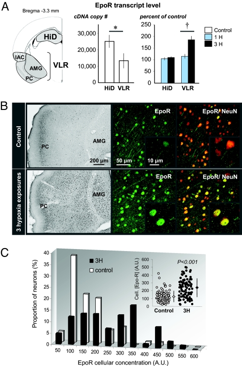
Erythropoietin receptor (EpoR) binding mediates neuroprotection by endogenous Epo or by exogenous recombinant human (rh)Epo. The level of EpoR gene expression may determine tissue responsiveness to Epo. Thus, harnessing the neuroprotective power of Epo requires an understanding of the Epo-EpoR system and its regulation. We tested the hypothesis that neuronal expression of EpoR is required to achieve optimal neuroprotection by Epo. The ventral limbic region (VLR) in the rat brain was used because we determined that its neurons express minimal EpoR under basal conditions, and they are highly sensitive to excitotoxic damage, such as occurs with pilocarpine-induced status epilepticus (Pilo-SE). We report that (i) EpoR expression is significantly elevated in nearly all VLR neurons when rats are subjected to 3 moderate hypoxic exposures, with each separated by a 4-day interval; (ii) synergistic induction of EpoR expression is achieved in the dorsal hippocampus and neocortex by the combination of hypoxia and exposure to an enriched environment, with minimal increased expression by either treatment alone; and (iii) rhEpo administered after Pilo-SE cannot rescue neurons in the VLR, unless neuronal induction of EpoR is elicited by hypoxia before Pilo-SE. This study thus demonstrates using environmental manipulations in normal rodents, the strict requirement for induction of EpoR expression in brain neurons to achieve optimal neuroprotection. Our results indicate that regulation of EpoR gene expression may facilitate the neuroprotective potential of rhEpo.


Conflict of interest statement

The authors declare no conflict of interest.

Figures

Fig. 1.
Fig. 1.
Repeated hypoxic exposures activate EpoR gene expression in the VLR. (A) EpoR transcript level measured by reverse transcription quantitative PCR (RT-qPCR) in the HiD and the VLR of control rats revealed that EpoR-mRNA level was lower in the VLR than that measured in the HiD (*, P < 0.05). When measured at reoxygenation time in rats subjected to either 1 (1H) or 3 (3H) hypoxic episodes, a significant increase in EpoR-mRNA level was found in the VLR after 3H only (†, P < 0.05, compared with 1H). All bars represent mean ± SEM (n = 4 in each group). (B) Three days after reoxygenation time in rats subjected to 3H, the number of cells in which EpoR was detected increased compared with controls, as shown on representative sections stained for chromogenic detection of EpoR. In sections processed for dual EpoR and NeuN immunofluorescent detection, all EpoR-positive cells appeared to be neurons (NeuN+), as illustrated in the IAC: EpoR is in green and NeuN in red. (C) After 3H, the increased number of cells detected by chromogenic immunohistochemistry in the IAC was associated with an increased EpoR cellular concentration index, which was determined as the intensity of the immunofluorescent labeling (n = 132 neurons in controls; n = 130 neurons after 3H). The illustration represents all neurons measured and the mean ± SD for each group.
Fig. 2.
Fig. 2.
Repeated hypoxic exposures induce Epo gene expression in the VLR. (A) Constitutive level of Epo transcript measured by RT-qPCR was similar in the HiD and the VLR. At reoxygenation time after 1H, Epo-mRNA level was significantly increased to the same extent in the 2 brain areas (P < 0.001 between control and 1H). However, at reoxygenation time after 3H, Epo-mRNA level was superinduced in the VLR only (†, P < 0.05; †††, P < 0.001 between 1H and 3H). All bars represent mean ± SEM (n = 4 in each group). (B) Three days after reoxygenation time in rats subjected to 3H, the number of cells in which Epo was detected increased compared with controls, as shown on representative sections stained for chromogenic detection of Epo. In sections processed for dual EpoR and NeuN immunofluorescent detection, all Epo-positive cells appeared to be neurons (NeuN+), as illustrated in the IAC: Epo is in green and NeuN in red. (C) After 3H, the increased number of cells detected by chromogenic immunohistochemistry in the IAC was associated with an increased Epo cellular concentration index, which was determined as the intensity of the immunofluorescent labeling (n = 141 neurons in controls; n = 153 neurons after 3H). Illustration represents all neurons measured and the mean ± SD for each group.
Fig. 3.
Fig. 3.
Environmental enrichment increases brain EpoR and Epo gene expression. (A) Level of EpoR transcript was measured in the NC, the HiD, the HiV, and the VLR of rats raised in SC or EC, and subjected or not to 3H. In 3H groups, tissues were collected at reoxygenation time of the last hypoxic exposure (*, P < 0.001, compared with SC; †, P < 0.001, compared with EC). (B) Three days after reoxygenation time in rats housed in EC and subjected to 3H, the number of cells in which EpoR was detected increased, compared with rats raised in SC. (C) Constitutive expression of Epo was increased in the HiD, the HiV, and the VLR in rats raised in EC compared with SC (*, P < 0.05). (D) Brain reactivity of Epo gene expression to 3H, measured at transcript level, was not affected by housing conditions. All bars represent mean ± SEM (n = 4 in each group).
Fig. 4.
Fig. 4.
Neuroprotective effects of rhEpo in the VLR are observed only in rats subjected to 3H after Pilo-SE. (A) Neuronal density in the VLR was measured at anatomical planes corresponding to interaural (IA) +6.44 and +5.40 mm, according to ref. . Because the anatomical plane itself had no effect, and no significant interaction was found between “anatomical plane” and “treatment condition,” results for neuronal density were collapsed over the anatomical plane factor. Neither 3H alone nor rhEpo alone had neuroprotective effects in the VLR after Pilo-SE. However, rhEpo administered in rats subjected to 3H significantly protected VLR neurons against neurodegeneration after Pilo-SE. (*, P < 0.001, compared with controls; †, P < 0.05) (n = 6 in each group). (B) Immunohistochemical detection of NeuN 6 days after Pilo-SE in the whole VLR indicates that the lesioned area (dotted lines) is considerably reduced only in rats subjected to 3H before Pilo-SE. (C) Enlarged illustrations of NeuN-immunohistochemical detection in the IAC. (D) Parenchymal uptake of rhEpo was greater in the VLR than that measured in the HiD (P < 0.001, ANOVA 2 in both control and Pilo-SE groups; factor 1 is “brain area,” factor 2 is “time after rhEpo injection”), both in controls and in rats subjected to Pilo-SE. Each bar represents the mean ± SEM (n = 3 in each group). ‡, time of termination.
Fig. 5.
Fig. 5.
Repeated hypoxic exposures do not alter IGF-1, Tpo, and TpoR transcript levels in the VLR. Controls and rats subjected to 3H were killed immediately after (R0), R1, and R2 days after the last hypoxic exposure. Levels of transcripts were measured by RT-qPCR. Each bar represents the mean ± SEM (n = 4 in each group).

References

    1. Brines M, Cerami A. Emerging biological roles for erythropoietin in the nervous system. Nat Rev Neurosci. 2005;6:484–494. - PubMed
    1. Maiese K, Chong ZZ, Li F, Shang YC. Erythropoietin: Elucidating new cellular targets that broaden therapeutic strategies. Prog Neurobiol. 2008;85:194–213. - PMC - PubMed
    1. Noguchi CT, Asavaritikrai P, Teng R, Jia Y. Role of erythropoietin in the brain. Crit Rev Oncol Hematol. 2007;64:159–171. - PMC - PubMed
    1. Ehrenreich H, et al. Erythropoietin therapy for acute stroke is both safe and beneficial. Mol Med. 2002;8:495–505. - PMC - PubMed
    1. Ehrenreich H, et al. Improvement of cognitive functions in chronic schizophrenic patients by recombinant human erythropoietin. Mol Psychiatry. 2007;12:206–220. - PubMed

Publication types

MeSH terms

LinkOut - more resources