

Retrieval of trophoblast cells from the cervical canal for prediction of abnormal pregnancy: a pilot study
- PMID: 19497946
- PMCID: PMC2727404
- DOI: 10.1093/humrep/dep206
Retrieval of trophoblast cells from the cervical canal for prediction of abnormal pregnancy: a pilot study
Abstract
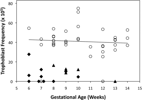
Background: Fetal cells are shed from the regressing chorionic villi and it is possible to retrieve extravillous cytotrophoblast cells by transcervical sampling. The abundance of trophoblast cells in transcervical samples suggests that this non-invasive approach could distinguish between normal and abnormal pregnancies, such as an ectopic pregnancy (EP) and blighted ovum (BO). We aim to identify and quantify fetal trophoblast cells in the cervical canal during the first trimester to assess their usefulness to predict an abnormal pregnancy.
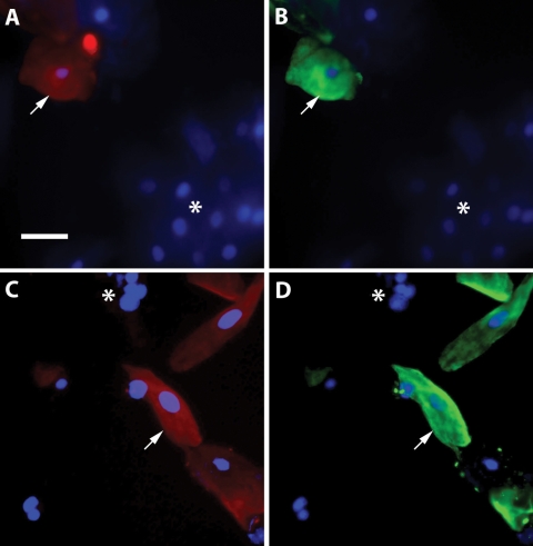
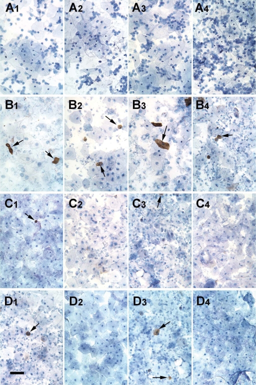
Methods: Patients, age 18-45, presenting with a normal intrauterine pregnancy (IUP; n = 37), diagnosis of EP (n = 10) or BO (n = 5) were enrolled for collection of transcervical specimens using a cytobrush and fixative rinse. Non-pregnant, nulliparous women (n = 7) were included as negative controls. Cells were cleared of mucus by acidification, prepared on microscope slides and labeled with a monoclonal antibody recognizing the trophoblast marker, human leukocyte antigen (HLA)-G. HLA-G positive and negative cells were counted to calculate the ratio of trophoblast cells to total cervical cells.
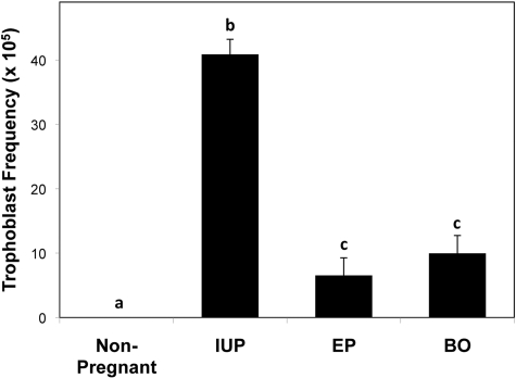
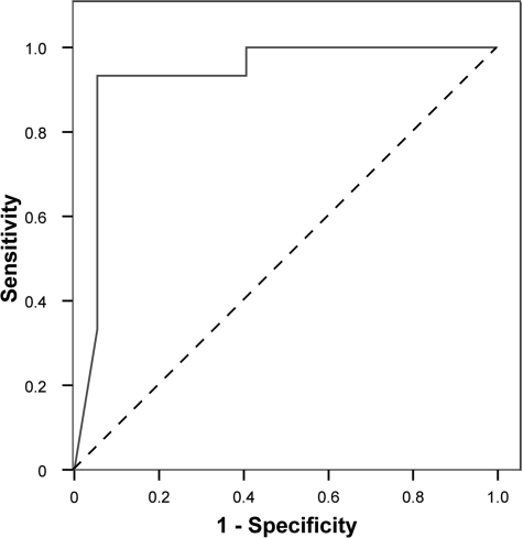
Results: Trophoblast cells were observed in 35/37 normal IUP, 6/10 EP and 4/5 BO specimens. The average frequency of HLA-G positive cells in the normal IUP cervical samples was approximately 1 in 2000, which was 4-fold higher than samples from patients with EP or BO (P < 0.001). Receiver operating characteristic analysis showed that EP and BO pregnancies were distinguishable from normal pregnancies with 93% sensitivity, 95% specificity, 97% positive predictive value and 87% negative predictive value.
Conclusions: This pilot study presents evidence that trophoblast cells can be reliably obtained and identified among cervical cells in the first trimester by immunohistochemical staining for HLA-G, and suggests for the first time that abnormal pregnancies may be predictable based on the abundance of trophoblast cells in the cervical canal.
Figures
Similar articles
-
HLA Class I protein expression in the human placenta.Early Pregnancy (Cherry Hill). 2001 Jan;5(1):67-9. Early Pregnancy (Cherry Hill). 2001. PMID: 11753519
-
Proteome-defined changes in cellular pathways for decidua and trophoblast tissues associated with location and viability of early-stage pregnancy.Reprod Biol Endocrinol. 2022 Feb 21;20(1):36. doi: 10.1186/s12958-022-00908-3. Reprod Biol Endocrinol. 2022. PMID: 35189928 Free PMC article.
-
Altered phenotype of HLA-G expressing trophoblast and decidual natural killer cells in pathological pregnancies.Hum Reprod. 2002 Apr;17(4):1072-80. doi: 10.1093/humrep/17.4.1072. Hum Reprod. 2002. PMID: 11925408
-
Transcervical retrieval of fetal cells in the practice of modern medicine: a review of the current literature and future direction.Fertil Steril. 2010 Apr;93(6):1725-30. doi: 10.1016/j.fertnstert.2009.11.022. Epub 2010 Jan 13. Fertil Steril. 2010. PMID: 20056202 Free PMC article. Review.
-
Immunological relationship between the mother and the fetus.Int Rev Immunol. 2002 Nov-Dec;21(6):471-95. doi: 10.1080/08830180215017. Int Rev Immunol. 2002. PMID: 12650238 Review.
Cited by
-
Endocervical trophoblast for interrogating the fetal genome and assessing pregnancy health at five weeks.Eur J Med Genet. 2019 Aug;62(8):103690. doi: 10.1016/j.ejmg.2019.103690. Epub 2019 Jun 18. Eur J Med Genet. 2019. PMID: 31226440 Free PMC article.
-
A Novel Paradigm for Non-Invasive Prenatal Genetic Screening: Trophoblast Retrieval and Isolation from the Cervix (TRIC).Diagnostics (Basel). 2023 Jul 30;13(15):2532. doi: 10.3390/diagnostics13152532. Diagnostics (Basel). 2023. PMID: 37568895 Free PMC article. Review.
-
Trophoblast retrieval and isolation from the cervix (TRIC) is unaffected by early gestational age or maternal obesity.Prenat Diagn. 2015 Dec;35(12):1218-22. doi: 10.1002/pd.4681. Epub 2015 Sep 20. Prenat Diagn. 2015. PMID: 26288006 Free PMC article.
-
Is cervical swab an efficient method for developing a new noninvasive prenatal diagnostic test for numerical and structural chromosome anomalies?Turk J Med Sci. 2021 Jun 28;51(3):1043-1048. doi: 10.3906/sag-2009-347. Turk J Med Sci. 2021. PMID: 33315353 Free PMC article.
-
Fetal Cell Based Prenatal Diagnosis: Perspectives on the Present and Future.J Clin Med. 2014 Sep 5;3(3):972-85. doi: 10.3390/jcm3030972. J Clin Med. 2014. PMID: 26237488 Free PMC article. Review.
References
-
- Adinolfi M, Sherlock J, Tutschek B, Halder A, Delhanty J, Rodeck C. Detection of fetal cells in transcervical samples and prenatal diagnosis of chromosomal abnormalities. Prenat Diagn. 1995;15:943–949. - PubMed
-
- Bulmer JN, Rodeck C, Adinolfi M. Immunohistochemical characterization of cells retrieved by transcervical sampling in early pregnancy. Prenat Diagn. 1995;15:1143–1153. - PubMed
-
- Bulmer JN, Cioni R, Bussani C, Cirigliano V, Sole F, Costa C, Garcia P, Adinolfi M. HLA-G positive trophoblastic cells in transcervical samples and their isolation and analysis by laser microdissection and QF-PCR. Prenat Diagn. 2003;23:34–39. - PubMed
-
- Bussani C, Cioni R, Scarselli B, Barciulli F, Bucciantini S, Simi P, Fogli A, Scarselli G. Strategies for the isolation and detection of fetal cells in transcervical samples. Prenat Diagn. 2002;22:1098–1101. - PubMed
-
- Bussani C, Cioni R, Mattei A, Fambrini M, Marchionni M, Scarselli G. Prenatal diagnosis of common aneuploidies in transcervical samples using quantitative fluorescent-PCR analysis. Mol Diagn Ther. 2007;11:117–121. - PubMed
Publication types
MeSH terms
Substances
Grants and funding
LinkOut - more resources
Full Text Sources
Other Literature Sources
Medical
Research Materials
