. 2009 Sep;30(8):1589-93.
doi: 10.3174/ajnr.A1602. Epub 2009 Jun 4.

Transcranial color-coded sonography successfully visualizes all intracranial parts of the internal carotid artery using the combined transtemporal axial and coronal approach

Affiliations

Transcranial color-coded sonography successfully visualizes all intracranial parts of the internal carotid artery using the combined transtemporal axial and coronal approach

J Eggers et al. AJNR Am J Neuroradiol. 2009 Sep.

Abstract

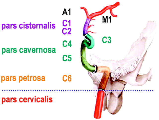
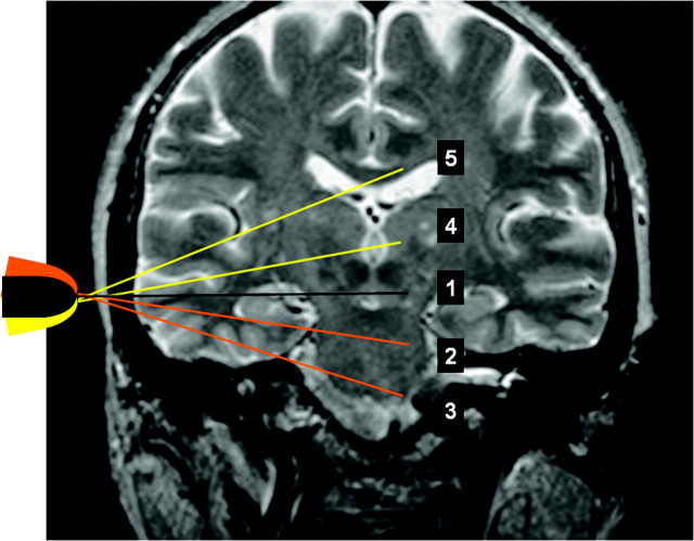
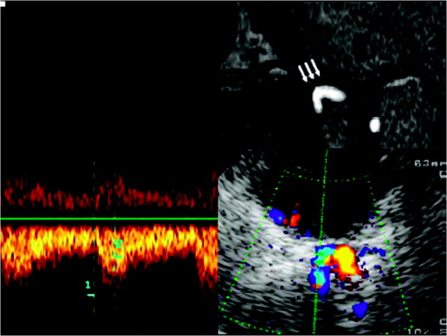
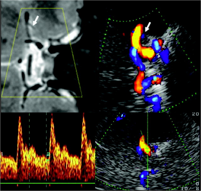
Background and purpose: Visualization of the intracranial internal carotid artery (ICA) with transcranial color-coded sonography (TCCS) by using the transtemporal coronal plane has been described previously. Because this approach is limited to the vertical running ICA segments, we investigated the feasibility of using TCCS to visualize all intracranial ICA segments by adding the transtemporal axial approach to the coronal plane.

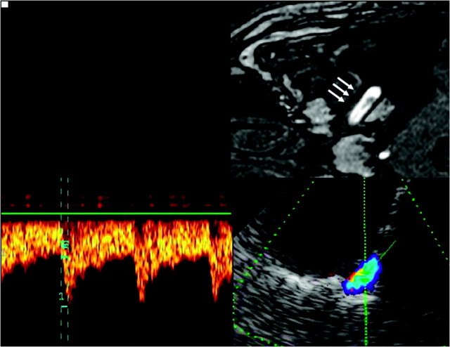
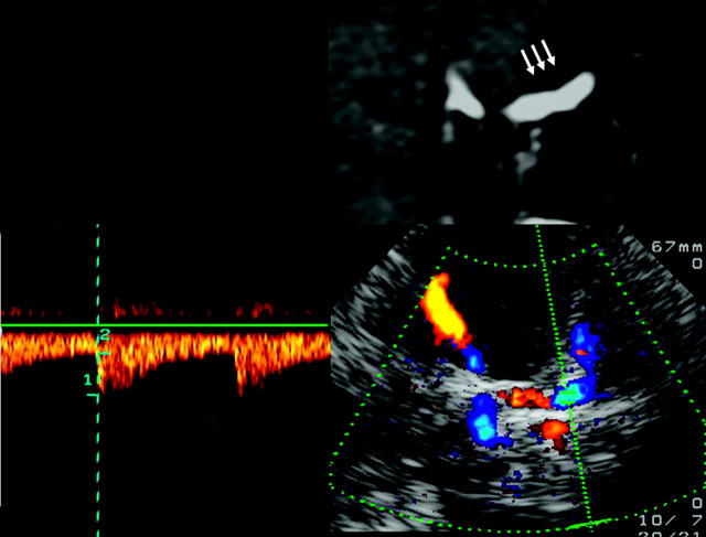
Materials and methods: Subjects with excellent transtemporal acoustic windows were examined by TCCS by using standardized axial and coronal planes. Identification rate, flow velocities, pulsatility and resistance indices, and length (as visible in color-coded power mode) were determined.

Results: A total of 120 intracranial ICAs from 60 subjects were investigated. By switching between the axial and coronal insonation planes, all intracranial segments of the ICA could be investigated in 100% of subjects-with the exception of the horizontal part of the petrosal ICA, which was identified in 96.7% of subjects.

Conclusions: TCCS becomes a reliable tool in investigating all parts of the intracranial ICA by adding the transtemporal axial approach to the coronal plane.


Figures

Fig 1.
Fig 1.
Sectioning of the ICA as used for sonographic examination, counting from the terminal to the proximal part to the intracranial ICA. Reprinted from Schünke et al with permission from Thieme Medical Publishers, Stuttgart, Germany. Note that this classification is different from the digital subtraction angiography classification, which names the cervical ICA, C1; the petrosal part, C2; and so on to the terminal ICA, C7 (Osborn).
Fig 2.
Fig 2.
Planes of transtemporal axial insonation: midbrain, upper pontine, lower pontine, thalamic, and cella media.
Fig 3.
Fig 3.
Assessment of the horizontal part of the C6 segment of the ICA, by using a transtemporal axial approach. Top: Time-of-flight MRA, axial source image, rotated 90° to correspond with the sonographic image. Horizontal part of the C6 segment (arrows). Bottom: TCCS, transtemporal approach, axial lower pontine plane. Right: Color-mode image demonstrates the horizontal part of the C6 segment. Left: Corresponding Doppler spectrum. Reprinted from Valdueza et al with permission from Thieme Medical Publishers, Stuttgart, Germany.
Fig 4.
Fig 4.
Assessment of the C5 segment of the ICA, by using a transtemporal coronal approach. Top: MR imaging, T1-weighted image, contrast-enhanced T1-weighted image, coronal plane, image rotated 90° to correspond with the sonographic image. Vertical part of the C5 segment (arrows). Bottom: TCCS, transtemporal approach, coronal plane. Right: Color-mode image demonstrating the C5 segment. Left: Corresponding Doppler spectrum. Reprinted from Valdueza et al with permission from Thieme Medical Publishers, Stuttgart, Germany.
Fig 5.
Fig 5.
Assessment of the carotid siphon by using a transtemporal axial approach. Top: Time of flight MRA, axial source image, rotated 90° to correspond with the sonographic imaging plane, C3–4 segments (arrows). Bottom: TCCS, transtemporal approach, axial upper pontine plane. Right: Color-mode image demonstrating the C-shaped part of the carotid siphon. Left: Corresponding Doppler spectrum. Reprinted from Valdueza et al with permission from Thieme Medical Publishers, Stuttgart, Germany.
Fig 6.
Fig 6.
Assessment of the terminal (C1–2) segments of the ICA, by using a transtemporal coronal approach. Top left: MR imaging, T2-weighted image, coronal plane, rotated 90° counter-clockwise. Flow voids in the C5 ICA, C1–2 ICA, M1 MCA, and A1 ACA segments of both sides. Top right: TCCS, transtemporal approach, anterior coronal plane. Corresponding color-mode image demonstrates blood flow in the distal C1–2 ICA segment, partly in the carotid siphon, in the A1 ACA segment, and in the proximal M1 MCA segment bilaterally. Note the red and blue color codes, indicating flow directions toward and away from the transducer. Bottom: Corresponding color-mode and Doppler spectrum of the C1–2 ICA segment. Reprinted from Valdueza et al with permission from Thieme Medical Publishers, Stuttgart, Germany.

Similar articles

Cited by

References

    1. Seidel G, Kaps M, Gerriets T. Potential and limitations of transcranial color-coded sonography in stroke patients. Stroke 1995; 26: 2061– 66 - PubMed
    1. Gerriets T, Postert T, Goertler M, et al. DIAS I: duplex-sonographic assessment of the cerebrovascular status in acute stroke—a useful tool for future stroke trials. Stroke 2000; 31: 2342– 45 - PubMed
    1. Gerriets T, Goertler M, Stolz E, et al. Feasibility and validity of transcranial duplex sonography for diagnosis in patients with acute stroke. J Neurol Neurosurg Psychiatry 2002; 72: 17– 20 - PMC - PubMed
    1. Hu HH, Luo CL, Sheng WY, et al. Transorbital color Doppler flow imaging of the carotid siphon and major arteries at the base of the brain. AJNR Am J Neuroradiol 1995; 16: 591– 98 - PMC - PubMed
    1. Jurgita V, Felix S, Thilo H, et al. Transcranial color-coded duplex sonography of the carotid siphon: the coronal plane approach. Clin Imaging 2002; 26: 81– 85 - PubMed

LinkOut - more resources