Review
. 2009 Jun;61(2):119-61.
doi: 10.1124/pr.109.001578. Epub 2009 Jun 4.

International Union of Basic and Clinical Pharmacology. LXXIII. Nomenclature for the formyl peptide receptor (FPR) family

Affiliations
Review

International Union of Basic and Clinical Pharmacology. LXXIII. Nomenclature for the formyl peptide receptor (FPR) family

Richard D Ye et al. Pharmacol Rev. 2009 Jun.

Abstract

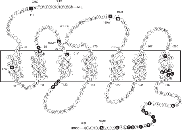
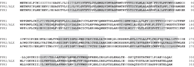
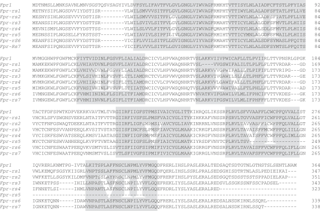
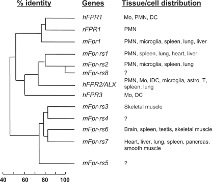
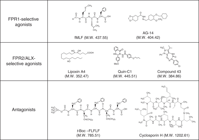
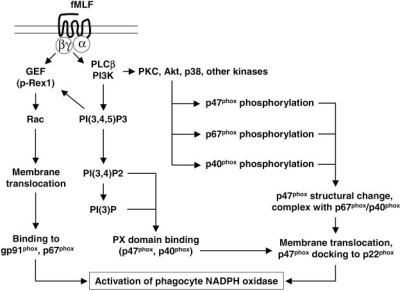
Formyl peptide receptors (FPRs) are a small group of seven-transmembrane domain, G protein-coupled receptors that are expressed mainly by mammalian phagocytic leukocytes and are known to be important in host defense and inflammation. The three human FPRs (FPR1, FPR2/ALX, and FPR3) share significant sequence homology and are encoded by clustered genes. Collectively, these receptors bind an extraordinarily numerous and structurally diverse group of agonistic ligands, including N-formyl and nonformyl peptides of different composition, that chemoattract and activate phagocytes. N-formyl peptides, which are encoded in nature only by bacterial and mitochondrial genes and result from obligatory initiation of bacterial and mitochondrial protein synthesis with N-formylmethionine, is the only ligand class common to all three human receptors. Surprisingly, the endogenous anti-inflammatory peptide annexin 1 and its N-terminal fragments also bind human FPR1 and FPR2/ALX, and the anti-inflammatory eicosanoid lipoxin A4 is an agonist at FPR2/ALX. In comparison, fewer agonists have been identified for FPR3, the third member in this receptor family. Structural and functional studies of the FPRs have produced important information for understanding the general pharmacological principles governing all leukocyte chemoattractant receptors. This article aims to provide an overview of the discovery and pharmacological characterization of FPRs, to introduce an International Union of Basic and Clinical Pharmacology (IUPHAR)-recommended nomenclature, and to discuss unmet challenges, including the mechanisms used by these receptors to bind diverse ligands and mediate different biological functions.


Figures

Fig. 1
Fig. 1
Predicted transmembrane disposition of the human FPR1. The protein sequence of the FPR-98 isoform (Leu110, Ala346) is shown (Boulay et al., 1990a). The transmembrane domains (TMs) are predicted based on hydrophobicity of the amino acid sequence and on similarities to the rhodopsin structure. The amino acids that form the boundaries of the transmembrane domains are numbered. One-letter amino acid code is used. The square blocks in reverce color represent positions at which amino acid substitutions result from polymorphisms, including amino acids 11 (Ile/Thr), 47 (Val/Ala), 101 (Leu/Val), 190 (Arg/Trp), 192 (Asn/Lys) and 346 (Ala/Glu). The circle blocks in reverse color indicate amino acids with known functions as follows. Arg84, Lys85, and Asp284 are critical for high-affinity binding of fMLF (Mills et al., 1998; Quehenberger et al., 1997). Asp122, Arg123, and Cys124 are the signature sequence for G protein interaction (DRY in many GPCRs). NPMLY in the TM7 are known signature sequence (NPXXY) for receptor internalization (Gripentrog et al., 2000; He et al., 2001). The 11 Ser and Thr residues in the cytoplasmic tail are potential phosphorylation sites for GRK2 and GRK3 (Prossnitz et al., 1995). CHO, carbohydrate, marks the identified and potential (in parenthesis) sites for N-glycosylation. The predicted disulfide bond between Cys98 and Cys176 is marked with double-line (=).
Fig. 2
Fig. 2
Alignment of the protein sequences of the human FPRs. The putative transmembrane domains (I to VII) are shaded. Sequence of the FPR-98 isoform is shown. Comparison of the three receptors has identified highly conserved regions, including most of TM-I and TM-II, and the short intracellular loop connecting TM-I and TM-II. The second intracellular loops from these receptors, known for G protein interaction, are nearly identical. TM-VII, including the NPXXY motif and a stretch of ∼25 amino acids extending toward the C-terminal tail, are also conserved among these receptors. Major differences are found in the extracellular domains between FPR1 and the other two receptors, especially in the amino termini (∼50% different), the second extracellular loops (56% different), and the third extracellular loops (∼50% different). The two putative N-glycosylation sites in the N-terminal domains are conserved among all three receptors. Most of the serines and threonines in the C-terminal tail, along with charged residues that constitute consensus GRK phosphorylation sites, are also conserved among these receptors.
Fig. 3
Fig. 3
Alignment of the predicted receptor sequence of the mouse Fpr genes. The putative transmembrane domains (TM-I–TM-VII) are shaded. Dashes indicate gaps in sequence created for alignment purposes. It is noteworthy that there is an eight-residue insertion in the N-terminal region of Fpr-1, just before TM-I. A three-residue insertion is found in the second extracellular loop in Fpr-1. The positively charged residues Arg84 and Lys85, found in human FPR1 and known for the interaction with fMLF, are missing from Fpr-1 and other mouse Fpr-related sequences. In its place are the noncharged residues Ser92 and Met93. The predicted Fpr-rs5 sequence is truncated at amino acid 246, resulting in a putative protein with only five TMs. Fpr-rs4 encodes a protein of 323 residues with a short C-terminal tail. In Fpr-rs1, there is a four-residue deletion in TM-IV, whereas the cloned mouse LXA4 receptor gene encodes a protein with the sequence of ARNV in its place. Polymorphisms exist in the Fpr-rs1 gene that result in amino acid substitutions at positions 3 (Thr/Ser), 8 (Pro/His), 13 (Asp/Glu), 16 (Ile/Val), 222 (Thr/Tyr), 236 (Phe/Ser), 296 (Ile/Met), and 318 (Gln/Pro) (Takano et al., 1997; Gao et al., 1998; Wang et al., 2002). The highest sequence identity (81%) is found between Fpr-rs1 and Fpr-rs2, and between Fpr-rs3 and Fpr-rs4.
Fig. 4
Fig. 4
Sequence homology between the FPR family members and their tissue distribution. The predicted protein sequences of the three human (h) FPR genes, the eight mouse (m) Fpr genes, and the rabbit (r) FPR1 gene were compared (Boulay et al., 1990a,b; Ye et al., 1993; Gao et al., 1998; Wang et al., 2002). Based on sequence homology, the hFPR1, mFpr1, and rFPR1 are in the same cluster. The mFpr-rs1, mFpr-rs2 (also termed mFpr2), and mFpr-rs8 are in another cluster closely related to hFPR2/ALX and hFPR3. The mFpr-rs3, mFpr-rs4, mFpr-rs6, and mFprrs7 (and, to a lesser extent, mFpr-rs5) are closely related based on their protein sequences (see Table 4 for sequence identity between the gene products). Note that some of these genes are not expressed in neutrophils and monocytes. The tissue expression profiles for mFpr-rs4, mFpr-rs5, and mFpr-rs8 have not been determined. Mo, monocytes; PMN, polymorphnuclear leukocytes; iDC, immature dendritic cells; astro, astrocytes; T, T lymphocytes.
Fig. 5
Fig. 5
Chemical structures of selected ligands for the formyl peptide receptors. Despite their abilities to bind to FPR1 and/or FPR2/ALX, these ligands have quite different structures. Of the ligands shown, t-Boc-FLFLF and CsH are antagonists and others are agonists. Note that the N-formyl group that defines agonistic activities in peptides such as fMLF is replaced with a bulky t-butyloxycarbonyl group that defines antagonistic activities in peptides such as t-Boc-FLFLF. LXA4, Quin-C1, and compound 43 are highly selective agonists for FPR2/ALX, whereas AG-14 and fMLF are selective for FPR1. t-Boc-FLFLF is selective for FPR1 at low concentrations but the selectivity is lost at high micromolar concentrations (e.g., 100 μM).
Fig. 6
Fig. 6
The fMLF-induced signaling events leading to the activation of phagocyte NADPH oxidase (Nox2). Depicted schematically are major signaling pathways activated by fMLF that results in NADPH oxidase activation in neutrophils. Upon activation of the Gαi proteins, the released Gβγ subunits trigger PI3K activation, resulting in the production PIP3 and its degradation products. The Gβγ and PIP3-mediated p-Rex1 activation is key to the conversion of GDP-bound Rac small GTPase to GTP-bound Rac, which translocates to membrane and associates with gp91phox.Gβγ is also responsible for PLCβ2 activation, leading to the production of the second mesengers diacyl glycerol and 1,4,5-inositol trisphosphate (IP3), which stimulate PKC activation. Isoforms of PKC (PKCδ, PKCξ, PKCβII, and PKCα), MAPK (p38, ERK), and Akt are known to catalyze the phosphorylation of p47phox in fMLF-stimulated neutrophils, prompting membrane translocation of the cytosolic factors. The PX domain in p47phox also facilitates its membrane association. Assembly of a membrane complex of NADPH oxidase is key to its conversion of molecular oxygen to superoxide. Omitted in this drawing is the phospholipase D (PLD) activation pathway, which is reported to contribute to fMLF-induced superoxide generation.

References

    1. Abo A, Pick E, Hall A, Totty N, Teahan CG, Segal AW. Activation of the NADPH oxidase involves the small GTP-binding protein p21rac1. Nature. 1991;353:668–670. - PubMed
    1. Agarwal S, Reynolds MA, Duckett LD, Suzuki JB. Altered free cytosolic calcium changes and neutrophil chemotaxis in patients with juvenile periodontitis. J Periodontal Res. 1989;24:149–154. - PubMed
    1. Ali H, Richardson RM, Haribabu B, Snyderman R. Chemoattractant receptor cross-desensitization. J Biol Chem. 1999;274:6027–6030. - PubMed
    1. Ali H, Richardson RM, Tomhave ED, Didsbury JR, Snyderman R. Differences in phosphorylation of formylpeptide and C5a chemoattractant receptors correlate with differences in desensitization. J Biol Chem. 1993;268:24247–24254. - PubMed
    1. Aliberti J, Hieny S, Reis e Sousa C, Serhan CN, Sher A. Lipoxin-mediated inhibition of IL-12 production by DCs: a mechanism for regulation of microbial immunity. Nat Immunol. 2002a;3:76–82. - PubMed

Publication types

MeSH terms

Substances

LinkOut - more resources