. 2009 Jun 5;324(5932):1330-4.
doi: 10.1126/science.1170905.

Mechanoenzymatic cleavage of the ultralarge vascular protein von Willebrand factor

Affiliations

Mechanoenzymatic cleavage of the ultralarge vascular protein von Willebrand factor

Xiaohui Zhang et al. Science. .

Abstract

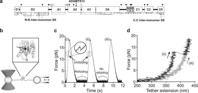
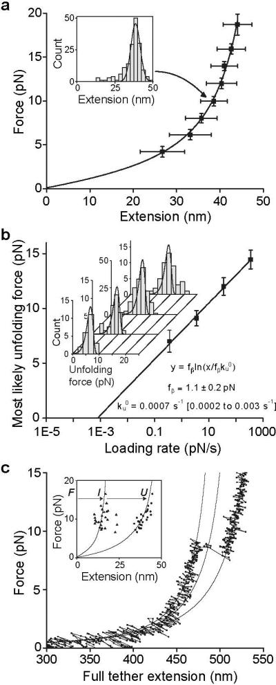
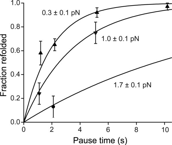
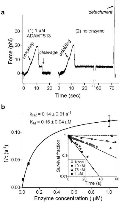
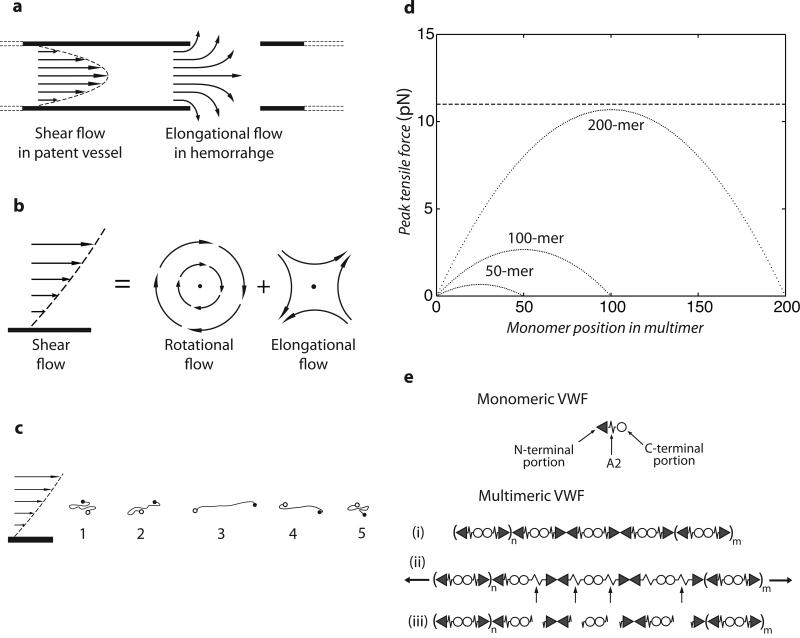
Von Willebrand factor (VWF) is secreted as ultralarge multimers that are cleaved in the A2 domain by the metalloprotease ADAMTS13 to give smaller multimers. Cleaved VWF is activated by hydrodynamic forces found in arteriolar bleeding to promote hemostasis, whereas uncleaved VWF is activated at lower, physiologic shear stresses and causes thrombosis. Single-molecule experiments demonstrate that elongational forces in the range experienced by VWF in the vasculature unfold the A2 domain, and only the unfolded A2 domain is cleaved by ADAMTS13. In shear flow, tensile force on a VWF multimer increases with the square of multimer length and is highest at the middle, providing an efficient mechanism for homeostatic regulation of VWF size distribution by force-induced A2 unfolding and cleavage by ADAMTS13, as well as providing a counterbalance for VWF-mediated platelet aggregation.


Figures

Figure 1
Figure 1. A2 domain unfolding and refolding with laser tweezers
a. Domain organization of VWF. Cysteines and disulfide bonds are shown beneath, and N- and O-linked sites above as filled and open lollipops, respectively. b. Experimental setup. A2 domain (enlarged in inset with ADAMTS13 cleavage site arrowed) is coupled to double-stranded DNA handles, which are bound through tags at their other ends to beads held by a laser trap and a translatable (arrows) micropipette. c. Force on a molecular tether during representative cycles of force increase, decrease, and clamping at a constant low level. d. Force-extension traces during force loading in cycles ii and iii from panel b.
Figure 2
Figure 2. Unfolding of the A2 domain
a. A2 domain force-extension data with an error-weighted least squares fit to the worm-like chain model (line) (14). Extension distances were sorted by unfolding force into 2 pN bins. A histogram of extensions for each bin (inset) was fit to a Gaussian (inset, solid line) to find peak extension, and force averaged for that bin. Uncertainty in extension is shown as the half width of the Gaussian fit and uncertainty in force is shown as one standard deviation. b. Unfolding force as a function of loading rate. Unfolding forces were binned by loading rate and plotted as histograms (inset). The peak of each histogram was plotted against the loading rate; uncertainty in y is shown as half of the bin width. A linear fit to the data (line) predicts the distributions of unfolding force (inset, lines), which agree well the histograms (inset). c. Representative force-extension trace for a tether pausing at an intermediate state, with three WLC curves (solid lines) representing DNA + folded A2, DNA + partially unfolded A2, and DNA + fully unfolded A2. Inset: Extensions of A2 to intermediate (I) and unfolded (U) lengths, fit to the WLC model (lines).
Figure 3
Figure 3. A2 domain refolding kinetics
Binary refolding events were binned by clamp force and time. Standard errors (bars) were calculated as (p*(1-p)/n)0.5, where p is fraction refolded and n is number of events. Overlaid on the data are the exponential curves predicted by maximum likelihood estimation (i.e. on the data without binning) using the τ∼ exp(f2) model.
Figure 4
Figure 4. Mechanoenzymatic cleavage of A2 by ADAMTS13
a. Representative traces showing cleavage in presence of enzyme ( panel 1) and no cleavage in absence of enzyme (panel 2) b. Enzyme kinetics. The hyperbolic dependence of catalytic rate on enzyme concentration was fit with the single-molecule Michaelis-Menten equation (21) (solid line). Data points and standard error were determined from single parameter exponential fits to the survival fraction as a function of time (inset).
Figure 5
Figure 5. Model for mechanoenzymatic cleavage of ULVWF in the circulation
a. Shear flow in a vessel and elongational flow at a site of bleeding. b. Shear flow may be represented as elongational flow superimposed on rotational flow (modified from (26)). c. Cartoon of VWF elongating, compressing, and tumbling in shear flow. d. Peak force as function of monomer position in a VWF multimer chain of 200, 100, or 50 monomers at 100 dyn/cm2. Dashed line shows the most likely unfolding force for the A2 domain at a loading rate of 25 pN/s. e. Schematic of VWF, with N-end as triangle, A2 as spring, and C-end as circle. Elongation results in unfolding of some A2 domains, some of which are cleaved (arrows). The resulting fragments are shown.

Comment in

References

    1. Sadler JE. Annu. Rev. Biochem. 1998;67:395. - PubMed
    1. Schneider SW, et al. Proc. Natl. Acad. Sci. U. S .A. 2007;104:7899. - PMC - PubMed
    1. Sadler JE. Annu. Rev. Med. 2005;56:173. - PubMed
    1. Batlle J, et al. Blood. 1987;70:173. - PubMed
    1. Tsai HM. Semin. Thromb. Haemost. 2003;29:479. - PubMed

Publication types