

Maternal endothelial progenitor colony-forming units with macrophage characteristics are reduced in preeclampsia
- PMID: 19498340
- PMCID: PMC2830891
- DOI: 10.1038/ajh.2009.101
Maternal endothelial progenitor colony-forming units with macrophage characteristics are reduced in preeclampsia
Abstract
Background: Endothelial progenitor cells (EPCs) provide paracrine support to the vascular endothelium and may also replace damaged or senescent endothelial cells. Low numbers of endothelial progenitor colony-forming units (CFU-ECs) in culture are a predictive biomarker of vascular disease. We hypothesized that the number of CFU-ECs derived from maternal blood are decreased in women with preeclampsia compared to normal pregnancy.
Methods: Primigravid women with singleton normal (n = 12) or preeclamptic (n = 12) pregnancies were studied during the third trimester. The culture assay was performed using a pre-plating step to eliminate mature endothelial cells and nonprogenitor cells; colonies per well were counted and further characterized.
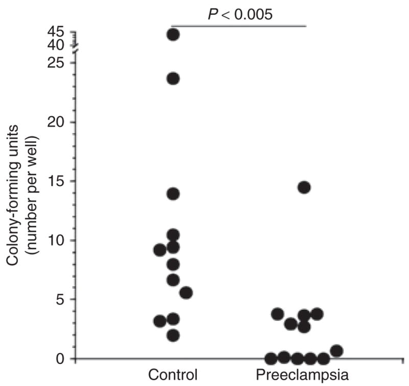
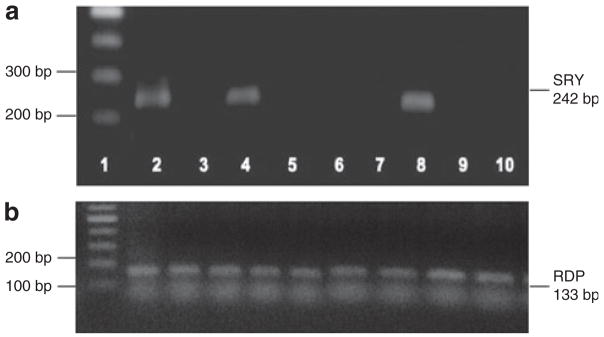
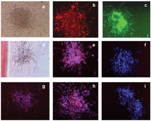
Results: Colony numbers were fourfold lower on average in preeclampsia compared to control samples (P < 0.005). A majority of the cells comprising individual colonies were positive for both endothelial (Ulex europaeus lectin staining and acetylated low-density lipoprotein (LDL) uptake) and monocyte/macrophage (CD45, CD14, CD115) characteristics. The SRY gene was detected in CFU-ECs derived from umbilical cord blood samples from male fetuses but not in CFU-ECs from peripheral blood of mothers with male fetuses. Maternal plasma concentrations of the antiangiogenic factor, soluble fms-like tyrosine kinase-1 (sFlt-1) were elevated (P < 0.0001) whereas placental growth factor (PlGF) was reduced (P < 0.01) in women with preeclampsia, but these factors did not correlate with CFU-EC counts.
Conclusions: CFU-ECs derived from culture of peripheral blood mononuclear cells, a correlate of cardiovascular risk in nonpregnancy populations, are rarified in women with preeclampsia compared to normal pregnancy. PCR analysis is consistent with a maternal origin of these cells.
Conflict of interest statement
Disclosure: The authors declared no conflict of interest.
Figures
References
-
- Roberts JM. Endothelial dysfunction in preeclampsia. Semin Reprod Endocrinol. 1998;16:5–15. - PubMed
-
- Ness RB, Sibai BM. Shared and disparate components of the pathophysiologies of fetal growth restriction and preeclampsia. Am J Obstet Gynecol. 2006;195:40–49. - PubMed
-
- Urbich C, Heeschen C, Aicher A, Dernbach E, Zeiher AM, Dimmeler S. Relevance of monocytic features for neovascularization capacity of circulating endothelial progenitor cells. Circulation. 2003;108:2511–2516. - PubMed
-
- Urbich C, Aicher A, Heeschen C, Dernbach E, Hofmann WK, Zeiher AM, Dimmeler S. Soluble factors released by endothelial progenitor cells promote migration of endothelial cells and cardiac resident progenitor cells. J Mol Cell Cardiol. 2005;39:733–742. - PubMed
-
- Hill JM, Zalos G, Halcox JP, Schenke WH, Waclawiw MA, Quyyumi AA, Finkel T. Circulating endothelial progenitor cells, vascular function, and cardiovascular risk. N Engl J Med. 2003;348:593–600. - PubMed
Publication types
MeSH terms
Substances
Grants and funding
LinkOut - more resources
Full Text Sources
Medical
Research Materials
Miscellaneous
