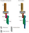
Tube Is an IRAK-4 homolog in a Toll pathway adapted for development and immunity
- PMID: 19498957
- PMCID: PMC2690232
- DOI: 10.1159/000200773
Tube Is an IRAK-4 homolog in a Toll pathway adapted for development and immunity
Abstract
Acting through the Pelle and IRAK family of protein kinases, Toll receptors mediate innate immune responses in animals ranging from insects to humans. In flies, the Toll pathway also functions in patterning of the syncytial embryo and requires Tube, a Drosophila -specific adaptor protein lacking a catalytic domain. Here we provide evidence that the Tube, Pelle, and IRAK proteins originated from a common ancestral gene. Following gene duplication, IRAK-4, Tube-like kinases, and Tube diverged from IRAK-1, Pelle, and related kinases. Remarkably, the function of Tube and Pelle in Drosophila embryos can be reconstituted in a chimera modeled on the predicted progenitor gene. In addition, a divergent property of downstream transcription factors was correlated with developmental function. Together, these studies reveal previously unrecognized parallels in Toll signaling in fly and human innate immunity and shed light on the evolution of pathway organization and function.
Keywords: Death domain; Drosophila; Evolution; Gene duplication; NF-κB; Signal transduction.
Figures


Similar articles
-
Pellino enhances innate immunity in Drosophila.Mech Dev. 2010 May-Jun;127(5-6):301-7. doi: 10.1016/j.mod.2010.01.004. Epub 2010 Feb 1. Mech Dev. 2010. PMID: 20117206
-
Phosphorylation modulates direct interactions between the Toll receptor, Pelle kinase and Tube.Development. 1998 Dec;125(23):4719-28. doi: 10.1242/dev.125.23.4719. Development. 1998. PMID: 9806920
-
IRAK-4: a novel member of the IRAK family with the properties of an IRAK-kinase.Proc Natl Acad Sci U S A. 2002 Apr 16;99(8):5567-72. doi: 10.1073/pnas.082100399. Proc Natl Acad Sci U S A. 2002. PMID: 11960013 Free PMC article.
-
Conventional and non-conventional Drosophila Toll signaling.Dev Comp Immunol. 2014 Jan;42(1):16-24. doi: 10.1016/j.dci.2013.04.011. Epub 2013 Apr 28. Dev Comp Immunol. 2014. PMID: 23632253 Free PMC article. Review.
-
Toll and Toll-like proteins: an ancient family of receptors signaling infection.Rev Immunogenet. 2000;2(3):294-304. Rev Immunogenet. 2000. PMID: 11256741 Review.
Cited by
-
Molecular evolution and structural features of IRAK family members.PLoS One. 2012;7(11):e49771. doi: 10.1371/journal.pone.0049771. Epub 2012 Nov 14. PLoS One. 2012. PMID: 23166766 Free PMC article.
-
Endocytosis is required for Toll signaling and shaping of the Dorsal/NF-kappaB morphogen gradient during Drosophila embryogenesis.Proc Natl Acad Sci U S A. 2010 Oct 19;107(42):18028-33. doi: 10.1073/pnas.1009157107. Epub 2010 Oct 4. Proc Natl Acad Sci U S A. 2010. PMID: 20921412 Free PMC article.
-
Structural biology: Immunity takes a heavy Toll.Nature. 2010 Jun 17;465(7300):882-3. doi: 10.1038/465882a. Nature. 2010. PMID: 20559378 No abstract available.
-
Immune-Related Genes in the Honey Bee Mite Varroa destructor (Acarina, Parasitidae).Insects. 2025 Mar 28;16(4):356. doi: 10.3390/insects16040356. Insects. 2025. PMID: 40332846 Free PMC article.
-
Comparative Genomics Reveals the Origins and Diversity of Arthropod Immune Systems.Mol Biol Evol. 2015 Aug;32(8):2111-29. doi: 10.1093/molbev/msv093. Epub 2015 Apr 22. Mol Biol Evol. 2015. PMID: 25908671 Free PMC article.
References
-
- Kawai T, Akira S. TLR signaling. Cell Death Differ. 2006;13:816–825. - PubMed
-
- Imler JL, Ferrandon D, Royet J, Reichhart JM, Hetru C, Hoffmann JA. Toll-dependent and Toll-independent immune responses in Drosophila. J Endotoxin Res. 2004;10:241–246. - PubMed
-
- Brennan CA, Anderson KV. Drosophila: the genetics of innate immune recognition and response. Annu Rev Immunol. 2004;22:457–483. - PubMed
-
- Pasare C, Medzhitov R. Toll-like receptors: linking innate and adaptive immunity. Adv Exp Med Biol. 2005;560:11–18. - PubMed
-
- Kim T, Kim YJ. Overview of innate immunity in Drosophila. J Biochem Mol Biol. 2005;38:121–127. - PubMed
Publication types
MeSH terms
Substances
Grants and funding
LinkOut - more resources
Full Text Sources
Other Literature Sources
Molecular Biology Databases

