

Evidence for ongoing brain injury in human immunodeficiency virus-positive patients treated with antiretroviral therapy
- PMID: 19499454
- PMCID: PMC2889153
- DOI: 10.1080/13550280902973960
Evidence for ongoing brain injury in human immunodeficiency virus-positive patients treated with antiretroviral therapy
Abstract
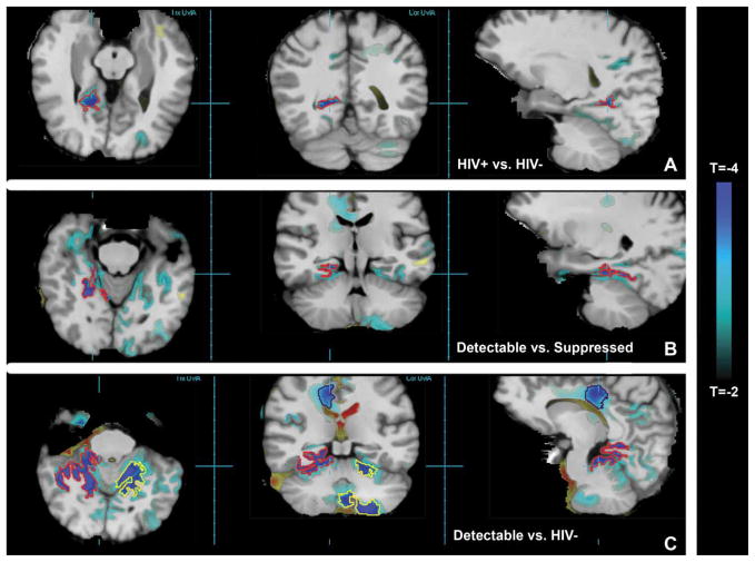
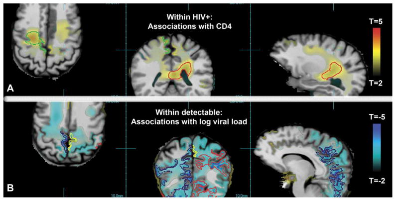
Treatment with antiretroviral therapy (ART) has greatly reduced the incidence of dementia. The goal of this longitudinal study was to determine if there are ongoing macrostructural brain changes in human immunodeficiency virus-positive (HIV + ) individuals treated with ART. To quantify brain structure, three-dimensional T1-weighted magnetic resonance imaging (MRI) scans were performed at baseline and again after 24 months in 39 HIV+ patients on ART and 30 HIV- controls. Longitudinal changes in brain volume were measured using tissue segmentation within regions of interest and deformation morphometry. Measured by tissue segmentation, HIV+ patients on ART had significantly (all P<.05) greater rates of white matter volume loss than HIV- control individuals. Compared with controls, the subgroup of HIV+ individuals on ART with viral suppression also had significantly greater rates of white matter volume loss. Deformation morphometry confirmed these results with more specific spatial localization. Deformation morphometry also detected greater rates of gray matter and white matter loss in the subgroup of HIV+ individuals with detectable viral loads. These results provide evidence of ongoing brain volume loss in HIV+ individuals on stable ART, possibly suggesting ongoing cerebral injury. The presence of continuing injury raises the possibility that HIV+ individuals-even in the presence of viral suppression in the periphery-are at greater risk for future cognitive impairments and dementia and possibly faster cognitive decline. Therefore, HIV+ individuals on ART should be monitored for cognitive decline, and treatments that reduce ongoing neurological injury should be considered.
Conflict of interest statement
Figures
References
-
- Agartz I, Brag S, Franck J, Hammarberg A, Okugawa G, Svinhufvud K, Bergman H. Mr volumetry during acute alcohol withdrawal and abstinence: a descriptive study. Alcohol Alcohol. 2003;38:71–78. - PubMed
-
- Ances BM, Roc AC, Wang J, Korczykowski M, Okawa J, Stern J, Kim J, Wolf R, Lawler K, Kolson DL, Detre JA. Caudate blood flow and volume are reduced in HIV+ neurocognitively impaired patients. Neurology. 2006;66:862–866. - PubMed
-
- Aylward EH, Henderer JD, McArthur JC, Brettschneider PD, Harris GJ, Barta PE, Pearlson GD. Reduced basal ganglia volume in HIV-1-associated dementia: results from quantitative neuroimaging. Neurology. 1993:2099–2104. - PubMed
Publication types
MeSH terms
Substances
Grants and funding
LinkOut - more resources
Full Text Sources
Other Literature Sources
Medical
