. 2009 Sep 15;125(6):1250-6.
doi: 10.1002/ijc.24509.

Human Merkel cell polyomavirus infection II. MCV is a common human infection that can be detected by conformational capsid epitope immunoassays

Affiliations

Human Merkel cell polyomavirus infection II. MCV is a common human infection that can be detected by conformational capsid epitope immunoassays

Yanis L Tolstov et al. Int J Cancer. .

Abstract

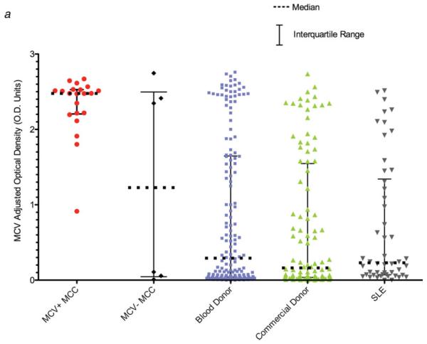
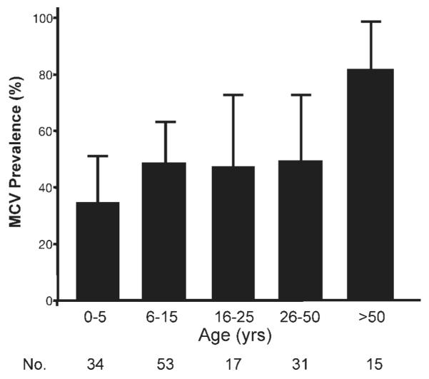
Merkel cell polyomavirus (MCV) is a newly-discovered human tumor virus found in approximately 80% of Merkel cell carcinoma (MCC). The rate of MCV infection among persons without MCC is unknown. We developed a MCV virus-like particle (VLP) enzyme-linked immunoassay (EIA) that does not cross-react with human BK or murine polyomaviruses. Peptide mapping of the MCV VP1 gene and immunoblotting with denatured MCV VLP are less sensitive than the MCV EIA in detecting MCV antibodies suggesting antibody reactivity in this assay primarily targets conformational but not linear epitopes. Among MCC patients, all 21 (100%) patients tested with MCV-positive tumors had high serum MCV IgG but not high MCV IgM levels. Only 3 of 6 (50%) MCC patients with MCV-negative tumors were positive for MCV antibodies. Sera from most adults, including 107 of 166 (64%) blood donors, 63 of 100 (63%) commercial donors and 37 of 50 (74%) systemic lupus erythematosus patients, show evidence for prior MCV exposure. Age-specific MCV prevalence was determined by examining a cross-sectional distribution of 150 Langerhans cell histiocytosis (an unrelated neoplasm) patient sera. MCV prevalence increases from 50% among children age 15 years or younger to 80% among persons older than 50 years. We did not find evidence for vertical transmission among infants. Although past exposure to MCV is common among all adult groups, MCC patients have a markedly elevated MCV IgG response compared with control patients. Our study demonstrates that MCV is a widespread but previously unrecognized human infection.


Figures

FIGURE 1
FIGURE 1
(a) MCV VP1 and VP2 proteins expressed in 293TT cells self-assemble into virus-like particles (VLP). This panel shows protein staining of gradient fractions from cell lysates expressing MCV strain 339 VP1 and VP2 proteins compared with murine polyomavirus VP1 and VP2, which are known to coassemble into VLP (Supporting Fig. 1). Each lane was loaded with 2.5 μl of fraction material, electrophoresed on a polyacrylamide gel and stained with SYPRO Ruby protein stain. Murine polyomavirus and MCV VP1 and VP2 proteins coassemble into high molecular weight complexes (fractions 7–9) with histone-associated DNA, which is characteristic for VLP production. (b) DNA concentrations, representing VLP encapsidation of cellular and plasmid DNA fragments, peak in high molecular weight gradient fractions 7–9 for both MCV and murine polyomavirus VLP. (c) Transmission electron microscopy of MCV VLP at ×50,000 magnification shows characteristic polyomaviral icosahedral capsid structures. Bar represents 100 nm.
FIGURE 2
FIGURE 2
(a) Anti-MCV VLP antibodies levels are significantly elevated in sera from MCV-positive MCC patients compared with other patient populations. IgG antibody levels against MCV were measured with an MCV VLP EIA for MCC patients, blood donors, commercial donors and SLE patients. Median MCV optical density values are indicated by solid horizontal lines, with bars representing interquartile values. Each sample was tested in duplicate, in a randomized and blinded fashion. Background subtraction in the absence of antigen was performed to obtain the adjusted optical density value for each serum. (b) Immune competition experiments for 4 MCC patient sera and 8 blood donor sera reactive on the MCV EIA. For the competitions, each serum was diluted to its EC50 concentration, preincubated with increasing amounts of either MCV (red) or BKV VLP, and then tested on the MCV EIA. MCV VLP but not BKV VLP compete for MCV reactivity among all of the sera. (c) No correlation is present between BKV and MCV IgG antibody responses in human sera measured by EIA (100 ng VLP protein per well). One hundred commercial donor samples were tested by BKV and MCV EIA at 1:500 dilution. The regression line between BKV and MCV antibody levels shows no correlation, with a slope that includes zero (95% CI, -0.15 to 0.03). Dotted lines represent 95% confidence bounds for the regression line.
FIGURE 2
FIGURE 2
(a) Anti-MCV VLP antibodies levels are significantly elevated in sera from MCV-positive MCC patients compared with other patient populations. IgG antibody levels against MCV were measured with an MCV VLP EIA for MCC patients, blood donors, commercial donors and SLE patients. Median MCV optical density values are indicated by solid horizontal lines, with bars representing interquartile values. Each sample was tested in duplicate, in a randomized and blinded fashion. Background subtraction in the absence of antigen was performed to obtain the adjusted optical density value for each serum. (b) Immune competition experiments for 4 MCC patient sera and 8 blood donor sera reactive on the MCV EIA. For the competitions, each serum was diluted to its EC50 concentration, preincubated with increasing amounts of either MCV (red) or BKV VLP, and then tested on the MCV EIA. MCV VLP but not BKV VLP compete for MCV reactivity among all of the sera. (c) No correlation is present between BKV and MCV IgG antibody responses in human sera measured by EIA (100 ng VLP protein per well). One hundred commercial donor samples were tested by BKV and MCV EIA at 1:500 dilution. The regression line between BKV and MCV antibody levels shows no correlation, with a slope that includes zero (95% CI, -0.15 to 0.03). Dotted lines represent 95% confidence bounds for the regression line.
FIGURE 3
FIGURE 3
Age-dependent prevalence of MCV antibodies among Langerhans cell histiocytosis (LCH) patients. Sera from a cross-sectional cohort of persons with a history of LCH were tested for IgG antibodies using the MCV EIA. Antibodies were absent from children 1 year and younger but increased in an age-dependent manner, reaching 80% prevalence among adults over age 50. Some children as young as 2 years old were strongly reactive on this assay.

References

    1. Dinh V, Feun L, Elgart G, Savaraj N. Merkel cell carcinomas. Hematol Oncol Clin North Am. 2007;21:527–44. - PubMed
    1. Engels EA, Frisch M, Goedert JJ, Biggar RJ, Miller RW. Merkel cell carcinoma and HIV infection. Lancet. 2002;359:497–8. - PubMed
    1. Feng H, Taylor JL, Benos PV, Newton R, Waddell K, Lucas SB, Chang Y, Moore PS. Human transcriptome subtraction by using short sequence tags to search for tumor viruses in conjunctival carcinoma. J Virol. 2007;81:11332–40. - PMC - PubMed
    1. Feng H, Shuda M, Chang Y, Moore PS. Clonal integration of a polyomavirus in human Merkel cell carcinoma. Science. 2008;319:1096–100. - PMC - PubMed
    1. Foulongne V, Kluger N, Dereure O, Brieu N, Guillot B, Segondy M. Merkel cell polyomavirus and Merkel cell carcinoma. Fr Emerg Infect Dis. 2008;14:1491–3. - PMC - PubMed

Publication types