. 2009 Sep;11(9):1365-81.
doi: 10.1111/j.1462-5822.2009.01337.x. Epub 2009 Jun 2.

Slc11a1 limits intracellular growth of Salmonella enterica sv. Typhimurium by promoting macrophage immune effector functions and impairing bacterial iron acquisition

Affiliations

Slc11a1 limits intracellular growth of Salmonella enterica sv. Typhimurium by promoting macrophage immune effector functions and impairing bacterial iron acquisition

Manfred Nairz et al. Cell Microbiol. 2009 Sep.

Abstract

The natural resistance-associated macrophage protein 1, Slc11a1, is a phagolysosomal transporter for protons and divalent ions including iron that confers host protection against diverse intracellular pathogens including Salmonella. We investigated and compared the regulation of iron homeostasis and immune function in RAW264.7 murine phagocytes stably transfected with non-functional Slc11a1 and functional Slc11a1 controls in response to an infection with Salmonella enterica serovar Typhimurium. We report that macrophages lacking functional Slc11a1 displayed an increased expression of transferrin receptor 1, resulting in enhanced acquisition of transferrin-bound iron. In contrast, cellular iron release mediated via ferroportin 1 was significantly lower in Salmonella-infected Slc11a1-negative macrophages in comparison with phagocytes bearing Slc11a1. Lack of Slc11a1 led to intracellular persistence of S. enterica serovar Typhimurium within macrophages, which was paralleled by a reduced formation of nitric oxide, tumour necrosis factor-alpha and interleukin-6 in Slc11a1-negative macrophages following Salmonella infection, whereas interleukin-10 production was increased. Moreover, Slc11a1-negative phagocytes exhibited higher cellular iron content, resulting in increased iron acquisition by intracellular Salmonella. Our observations indicate a bifunctional role for Slc11a1 within phagocytes. Slc11a restricts iron availability, which first augments pro-inflammatory macrophage effector functions and second concomitantly limits microbial iron access.


Figures

Figure 1
Figure 1. Effects of Slc11a1 on iron acquisition and iron release by Salmonella-infected macrophages
Slc11a1-non-functional RAW-21 cells (light grey bars) and Slc11a1-functional RAW-37 cells (dark grey bars) were infected with S. Typhimurium (S. tm.) at a MOI of 10 and stimulated with 50 U/ml IFN-γ for 24 hours. TfR1 and Fpn1 mRNA levels (Figures 1A and 1C, respectively) were determined by qPCR. Values were corrected for the amount of 18S ribosomal RNA, which was determined in parallel. Results are shown as relative differences of this ratio in comparison to unstimulated Slc11a1-expressing RAW-37 control macrophages (=1.0). Data are expressed as mean ± SD of five independent experiments. The uptake of TBI as well as iron release (depicted in Figures 1B and 1D and, respectively) were determined as described in ‘Experimental procedures’. Data are shown as mean ± SD of five independent experiments performed in duplicates and are expressed as fold change in the relative iron uptake/release in comparison to RAW-37 controls (=1.0). * P < 0.05 for infected/stimulated RAW-21 macrophages in comparison to RAW-21 controls; ° P < 0.05 for infected/stimulated RAW-37 in comparison to untreated RAW-37 cells; # P < 0.05 for the comparison of RAW-37 and RAW-21 cells subjected to the same treatment.
Figure 2
Figure 2. Regulation of TfR1 and Fpn1 protein levels in Salmonella-infected RAW-21 and RAW-37 cells
Slc11a1-non-expressing RAW-21 cells and Slc11a1-expressing RAW-37 cells were infected with Salmonella and stimulated with IFN-γ exactly as described in the legend to Figure 1. Whole cell lysates were analysed by immunoblotting using specific antibodies to TfR1 (upper panel) and Fpn1 (middle panel). Equal loading of protein extracts was confirmed by reprobing membranes with an anti-Actin antibody (lower panel). One of four representative immunoblot experiments is shown.
Figure 3
Figure 3. Effects of Slc11a1 on ferritin expression and on intracellular iron content in Salmonella-infected macrophages
RAW-21 and RAW-37 cells were infected with Salmonella and activated with IFN-γ exactly as described in the legend to Figure 1. The mRNA levels of H-ferritin were determined by qPCR (Figure 3A). Results are shown as relative differences of this ratio in comparison to the unstimulated control macrophages (=1.0). The total cellular iron content was determined by means of atomic absorption spectrometry and normalized for the protein content (Figure 3B). Data are expressed as means ± SD of five independent experiments. Statistically significant differences are indicated as described in the legend to figure 1.
Figure 4
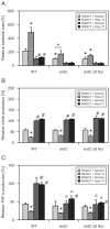
Figure 4. Impact of Slc11a1 on macrophage immune effector functions after IFN-γ stimulation
RAW-21 and RAW-37 cells were stimulated with IFN-γ and/or infected with Salmonella for 24 hours. Culture supernatants were analyzed for RNS (as determined by measuring nitrite levels, Figure 4A), TNF-α (Figure 4B), IL-6 (Figure 4C) and IL-10 (Figure 4D) as described in ‘Experimental procedures’. Data are shown as mean ± SD of four to five independent experiments. Statistically significant differences are indicated as described in the legend to figure 1.
Figure 5
Figure 5. Impact of Slc11a1 on the intracellular survival of Salmonella
RAW-21 and RAW-37 cells were infected with Salmonella for the indicated periods (Figure 5A) or infected for 24 hours and activated with IFN-γ (Figure 5B). Bacterial load was determined by selective plating as described in ‘Experimental procedures’.
Figure 6
Figure 6. Impact of Slc11a1 on macrophage immune effector functions in the presence of exogenous iron perturbations
In additional experiments, macrophages were infected with Salmonella and exposed to serial concentrations of human holo-transferrin. Bacterial load was determined by selective plating (Figure 6A). Cell culture supernatants were analyzed for the formation of nitrite, TNF-α, IL-10 and IL-6 as described (Figures 6B, C, D and E, respectively). Statistically significant differences are indicated as described
Figure 7
Figure 7. Contributions of Fpn1-mediated iron export and of TBI-uptake to Slc11a1-dependent macrophage functions
RAW-21 and RAW-37 cells were pre-treated for 8 hours with FeSO4, DFO, synthetic Hamp, a blocking α-TfR1 antibody or the appropriate control. Subsequently, cells were infected with Salmonella for 12 hours. Intracellular bacteria were enumerated (Figure 7A) and concentrations of TNF-α (Figure 7B) and of nitrite (Figure 7C) in supernatants were measured by a specific ELISA or the Griess reaction, respectively.
Figure 8
Figure 8. Limitation of iron availability for intramacrophage Salmonella in the presence of Slc11a1
The acquisition of NTBI (Figure 8A) and TBI (Figure 8B) by intramacrophage bacilli was determined as described in ‘Experimental procedures’ using S. Typhimurium engulfed by Slc11a1-non-functional RAW-21 in comparison to Slc11a1-functional RAW-37 phagocytes. Data are shown as means ± SD of five independent experiments and normalized for the number of bacteria as determined by selective plating.
Figure 9
Figure 9. Antibacterial effects in RAW-21 and RAW-37 cells infected with iron-uptake mutant Salmonella
RAW-21 and RAW-37 phagocytes were infected with S. Typhimurium wild-type strain as well as with isogenic entC and entC sit feo mutant derivatives and treated with PBS or 12.5 μg/ml holo-transferrin. The intracellular survival of bacteria was enumerated after 20 hours, and data are presented as relative values in comparison to solvent-treated RAW-37 cells infected with wild-type Salmonella (Figure 9A). Culture supernatants were analyzed for the concentrations of TNF-α (Figure 9B) and nitrite (9C).

Similar articles

Cited by

References

    1. Ables GP, Takamatsu D, Noma H, El-Shazly S, Jin HK, Taniguchi T, et al. The roles of Nramp1 and Tnfa genes in nitric oxide production and their effect on the growth of Salmonella typhimurium in macrophages from Nramp1 congenic and tumor necrosis factor-alpha−/− mice. J Interferon Cytokine Res. 2001;21:53–62. - PubMed
    1. Atkinson PG, Barton CH. High level expression of Nramp1G169 in RAW264.7 cell transfectants: analysis of intracellular iron transport. Immunology. 1999;96:656–662. - PMC - PubMed
    1. Atkinson PG, Blackwell JM, Barton CH. Nramp1 locus encodes a 65 kDa interferon-gamma-inducible protein in murine macrophages. Biochem J. 1997;325 ( Pt 3):779–786. - PMC - PubMed
    1. Barton CH, Biggs TE, Baker ST, Bowen H, Atkinson PG. Nramp1: a link between intracellular iron transport and innate resistance to intracellular pathogens. J Leukoc Biol. 1999;66:757–762. - PubMed
    1. Blackwell JM, Goswami T, Evans CA, Sibthorpe D, Papo N, White JK, et al. SLC11A1 (formerly NRAMP1) and disease resistance. Cell Microbiol. 2001;3:773–784. - PMC - PubMed

Publication types

MeSH terms