. 2009 Jun 5:7:60.
doi: 10.1186/1477-7827-7-60.

Hormone-regulated expression and distribution of versican in mouse uterine tissues

Affiliations

Hormone-regulated expression and distribution of versican in mouse uterine tissues

Renato M Salgado et al. Reprod Biol Endocrinol. .

Abstract

Background: Remodeling of the extracellular matrix is one of the most striking features observed in the uterus during the estrous cycle and after hormone replacement. Versican (VER) is a hyaluronan-binding proteoglycan that undergoes RNA alternative splicing, generating four distinct isoforms. This study analyzed the synthesis and distribution of VER in mouse uterine tissues during the estrous cycle, in ovariectomized (OVX) animals and after 17beta-estradiol (E2) and medroxyprogesterone (MPA) treatments, either alone or in combination.

Methods: Uteri from mice in all phases of the estrous cycle, and animals subjected to ovariectomy and hormone replacement were collected for immunoperoxidase staining for versican, as well as PCR and quantitative Real Time PCR.

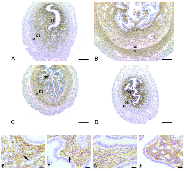
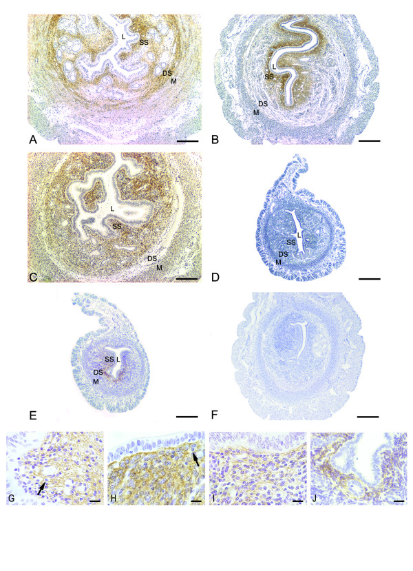
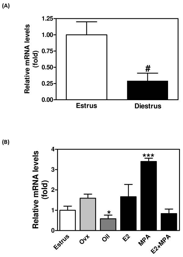
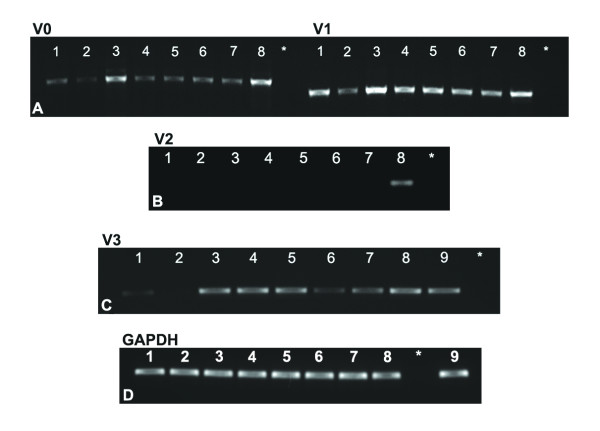
Results: In diestrus and proestrus, VER was exclusively expressed in the endometrial stroma. In estrus and metaestrus, VER was present in both endometrial stroma and myometrium. In OVX mice, VER immunoreaction was abolished in all uterine tissues. VER expression was restored by E2, MPA and E2+MPA treatments. Real Time PCR analysis showed that VER expression increases considerably in the MPA-treated group. Analysis of mRNA identified isoforms V0, V1 and V3 in the mouse uterus.

Conclusion: These results show that the expression of versican in uterine tissues is modulated by ovarian steroid hormones, in a tissue-specific manner. VER is induced in the myometrium exclusively by E2, whereas MPA induces VER deposition only in the endometrial stroma.


Figures

Figure 1
Figure 1
Immunoperoxidase for versican. (A) proestrus and (D) diestrus: the immunoreaction is seen as a dense network in the endometrial stroma, but absent in the myometrium. In both groups, the immunolabeling is stronger in the superficial stroma (SS) and only traces are observed in the interface between deep stroma (DS) and myometrium (M); (B) estrus and (C) metaestrous: the immunoreaction is observed in both superficial (SS) and deep stroma (DS). In these phases versican is also immunodetected in the internal layer of the myometrium (M); L: Lumen; SS: Superficial Stroma; DS: Deep Stroma; M: Myometrium. Scale bar: 200 μm. Higher Magnification micrographs show the localization of VER inside and outside the cells in proestrus (E), estrus (F) and diestrus (H), and mostly outside the cells in metaestrus (G). The reaction is absent from immune cells cytoplasm (arrows). Scale bar: 20 μm.
Figure 2
Figure 2
Immunoperoxidase for versican. (A) E2 treatment: strong immunoreaction is present in the whole endometrial stroma, except in areas of edema. The reaction can be observed as a network of delicate filaments. The immunolabeling is also observed at the interface between the deep stroma (DS) and the myometrium (M), as well as in the myometrial internal layer; (B) MPA treatment: versican distribution is seen as a dense brownish network in the extracellular spaces, exclusively in the superficial stroma (SS); (C) E2+MPA treatment: the immunoreaction is observed as a network of thin filaments in the whole endometrial stroma, except at the interface between the deep stroma (DS) and the myometrium (M). (D) ovariectomized group: immunoreaction is absent from the uterine tissues; (E) oil control group: immunoreaction is seen underneath the luminal epithelium of the antimesometrial stroma. The negative control shows no immunoreaction (F). L: Lumen; SS: Superficial Stroma; DS: Deep Stroma; M: Myometrium. Scale bar: 200 μm. Higher Magnification micrographs show the localization of VER inside and outside the cells in the MPA (H) and oil (J) groups, and mostly outside the cells in the E2 (G) and E2+MPA (I) groups. The reaction is absent from immune cells cytoplasm (arrows). Scale bar: 20 μm.
Figure 3
Figure 3
Relative mRNA expression of versican in the mouse uterus. The mRNA expression was analyzed by real-time PCR using primers common to all splice variants, and relative gene expression determined by designating estrus to 1. (A) versican mRNA in estrus and diestrus; (B) versican mRNA after ovariectomy and hormone replacement. Values represent the mean + SE of determinations on three independent tissue preparations. # p < 0.05 vs. estrus by Student t-test; *p < 0.05 vs. ovx and E2; ** p < 0.001 vs. all by ANOVA.
Figure 4
Figure 4
Expression of versican isoforms. Gel analysis of products generated by RT-PCR using specific primer sets for versican V0, V1, V2 and V3. Note the presence of V0 (left panel), V1 (right panel) (A) and V3 (C), but the absence of V2 (B). GAPDH internal control shows uniform expression in all groups studied (D). Total RNA isolated from whole mouse uterus from all groups studied (n = 5). 1- Diestrus; 2- Estrus; 3- E2; 4- MPA; 5- E2+MPA; 6- ovx; 7- oil; 8- UMRR (Universal Mouse Reference RNA); 9- ovary; * – negative control.

References

    1. Allen E. The oestrous cycle in the mouse. Am J Anat. 1927;30:297–371. doi: 10.1002/aja.1000300303. - DOI
    1. Martin L, Finn CA. Hormone secretion during early pregnancy in the mouse. J Endocrinol. 1969;45:57–65. doi: 10.1677/joe.0.0450057. - DOI - PubMed
    1. Parandoosh Z, Crombie DL, Tetzke TA, Hayes JS, Heap RB, Wang M-W. Progesterone and oestrogen receptors in the decidualized mouse uterus and effects of different types of anti-progesterone treatment. J Reprod Fertil. 1995;105:215–220. - PubMed
    1. Lydon JP, DeMayo FJ, Funk CR, Mani SK, Hughes AR, Montgomery CA, Jr, Shyamala G, Conneely OM, O'Malley BW. Mice lacking progesterone receptor exhibit pleiotropic reproductive abnormalities. Genes & Development. 1995;9:2266–2278. doi: 10.1101/gad.9.18.2266. - DOI - PubMed
    1. Weihua Z, Saji S, Sirpa M, Cheng G, Jensen EV, Warner M, Gustafsson J. Estrogen receptor (ER) β, a modulator of ERα in the uterus. Procl Natl Acad Sci USA. 2000;97:5936–5941. doi: 10.1073/pnas.97.11.5936. - DOI - PMC - PubMed

Publication types

LinkOut - more resources