. 2009 Jun 7:8:122.
doi: 10.1186/1475-2875-8-122.

Human T cell recognition of the blood stage antigen Plasmodium hypoxanthine guanine xanthine phosphoribosyl transferase (HGXPRT) in acute malaria

Affiliations

Human T cell recognition of the blood stage antigen Plasmodium hypoxanthine guanine xanthine phosphoribosyl transferase (HGXPRT) in acute malaria

Tonia Woodberry et al. Malar J. .

Abstract

Background: The Plasmodium purine salvage enzyme, hypoxanthine guanine xanthine phosphoribosyl transferase (HGXPRT) can protect mice against Plasmodium yoelii pRBC challenge in a T cell-dependent manner and has, therefore, been proposed as a novel vaccine candidate. It is not known whether natural exposure to Plasmodium falciparum stimulates HGXPRT T cell reactivity in humans.

Methods: PBMC and plasma collected from malaria-exposed Indonesians during infection and 7-28 days after anti-malarial therapy, were assessed for HGXPRT recognition using CFSE proliferation, IFNgamma ELISPOT assay and ELISA.

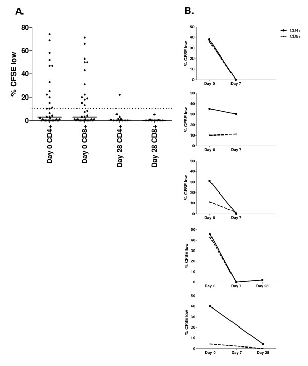
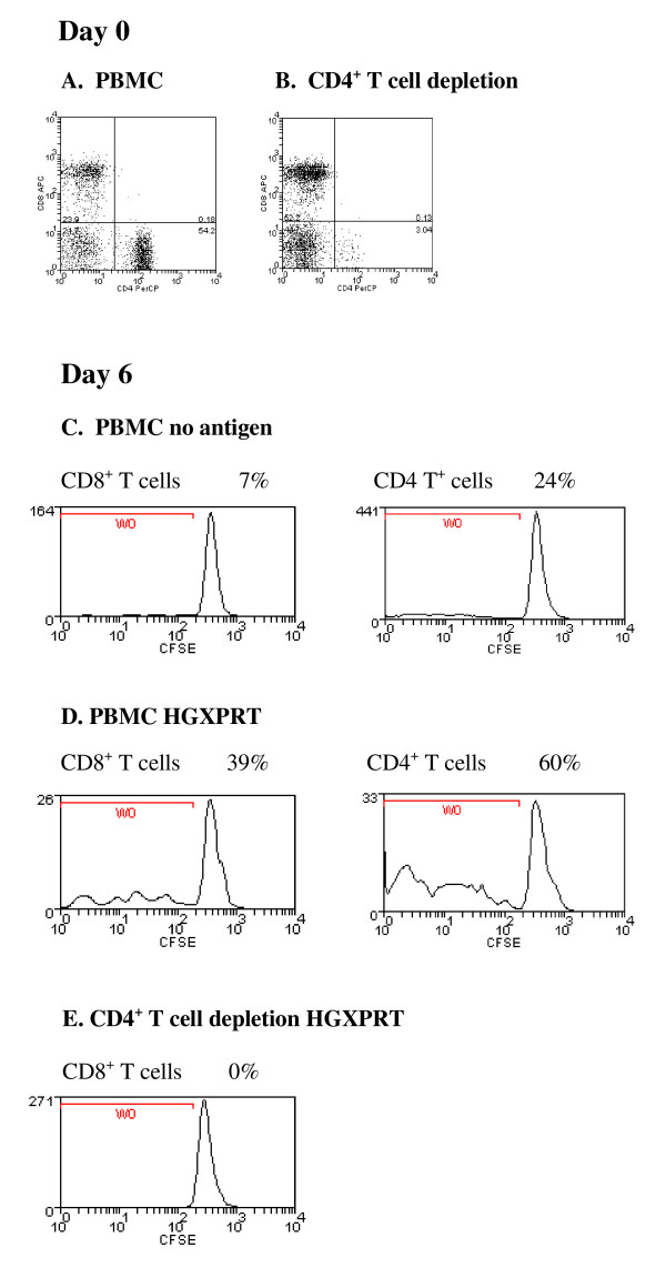
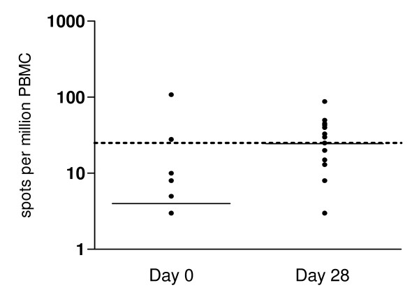
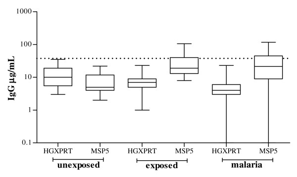
Results: HGXPRT-specific T cell proliferation was found in 44% of patients during acute infection; in 80% of responders both CD4+ and CD8+ T cell subsets proliferated. Antigen-specific T cell proliferation was largely lost within 28 days of parasite clearance. HGXPRT-specific IFN-gamma production was more frequent 28 days after treatment than during acute infection. HGXPRT-specific plasma IgG was undetectable even in individuals exposed to malaria for at least two years.

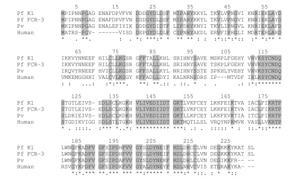
Conclusion: The prevalence of acute proliferative and convalescent IFNgamma responses to HGXPRT demonstrates cellular immunogenicity in humans. Further studies to determine minimal HGXPRT epitopes, the specificity of responses for Plasmodia and associations with protection are required. Frequent and robust T cell proliferation, high sequence conservation among Plasmodium species and absent IgG responses distinguish HGXPRT from other malaria antigens.


Figures

Figure 1
Figure 1
Lymphocyte proliferation to HGXPRT in acute and convalescent malaria. CD4+ and CD8+ T cell division following PBMC stimulation with HGXPRT expressed as the percentage of CFSE dim cells following background subtraction. A. PBMC response in 34 acute (day 0) and 12 convalescent (day 28) subjects. The proportion with proliferation at day 28 was significantly less than at day 0 (p = 0.035). No HGXPRT proliferation was detected in 15 malaria unexposed controls. The horizontal solid line represents the group median and the dotted line the background cut-off for positive responses. B. Longitudinal HGXPRT responses in 5 subjects during acute malaria (day 0) and 7 and 28 days after drug treatment.
Figure 2
Figure 2
Loss of proliferation to HGXPRT following CD4+ T cell depletion. Detection of HGXPRT-specific CD4+ or CD8+ T cells from a representative patient with acute falciparum malaria; (A) before and (B) after CD4+ T cell depletion. Day 6 CFSE CD8+ and CD4+ T cell proliferative responses in response to (C) no antigen and (D) HGXPRT before and (E) after CD4+ T cell depletion. The percentages represent the proportion of CFSE low CD4+ and CD8+ T cells in culture. A similar result was observed in two other subjects.
Figure 3
Figure 3
Plasmodium and Homo sapiens HG(X)PRT sequence alignment. Sections highlighted in grey, and marked with an asterix [*] show identical amino acids in the four sequences. A colon [:] indicates highly conserved amino acids and a single dot [.] indicates reasonable conservation between the Plasmodial (accession number; XP_001614435, P07833, P20035) and human HG(X)PRT (NP_000185) sequences.
Figure 4
Figure 4
Lymphocyte IFN-γ secretion to HGXPRT in acute and convalescent malaria: Ex-vivo ELISPOT detection of IFN-γ secretion following HGXPRT stimulation. PBMC responses in 12 acute (day 0) and 19 convalescent (day 28) subjects are shown after background subtraction. Spot forming cells were significantly more numerous during convalescence (p = 0.04). The horizontal solid line represents the group median and the dotted line the background cut-off for positive responses.
Figure 5
Figure 5
Antibody responses to P. falciparum HGXPRT and MSP5. Plasma IgG responses in 37 unexposed donors, 85 malaria exposed asymptomatic controls and 80 people with acute malaria (72 tested for MSP5 recognition). The solid line represents the group median and the dotted line the cut-off for positive responses as defined in the methods.

References

    1. Snow RW, Guerra CA, Noor AM, Myint HY, Hay SI. The global distribution of clinical episodes of Plasmodium falciparum malaria. Nature. 2005;434:214–217. doi: 10.1038/nature03342. - DOI - PMC - PubMed
    1. Clyde DF, Most H, McCarthy VC, Vanderberg JP. Immunization of man against sporozite-induced falciparum malaria. Am J Med Sci. 1973;266:169–177. doi: 10.1097/00000441-197309000-00002. - DOI - PubMed
    1. Hoffman SL, Goh LM, Luke TC, Schneider I, Le TP, Doolan DL, Sacci J, de la Vega P, Dowler M, Paul C, et al. Protection of humans against malaria by immunization with radiation-attenuated Plasmodium falciparum sporozoites. J Infect Dis. 2002;185:1155–1164. doi: 10.1086/339409. - DOI - PubMed
    1. Pombo DJ, Lawrence G, Hirunpetcharat C, Rzepczyk C, Bryden M, Cloonan N, Anderson K, Mahakunkijcharoen Y, Martin LB, Wilson D, et al. Immunity to malaria after administration of ultra-low doses of red cells infected with Plasmodium falciparum. Lancet. 2002;360:610–617. doi: 10.1016/S0140-6736(02)09784-2. - DOI - PubMed
    1. Edstein MD, Kotecka BM, Anderson KL, Pombo DJ, Kyle DE, Rieckmann KH, Good MF. Lengthy antimalarial activity of atovaquone in human plasma following atovaquone-proguanil administration. Antimicrob Agents Chemother. 2005;49:4421–4422. doi: 10.1128/AAC.49.10.4421-4422.2005. - DOI - PMC - PubMed

Publication types

MeSH terms

Substances