. 2009 Aug 14;391(2):341-58.
doi: 10.1016/j.jmb.2009.05.078. Epub 2009 Jun 3.

Two structurally independent domains of E. coli NusG create regulatory plasticity via distinct interactions with RNA polymerase and regulators

Affiliations

Two structurally independent domains of E. coli NusG create regulatory plasticity via distinct interactions with RNA polymerase and regulators

Rachel Anne Mooney et al. J Mol Biol. .

Abstract

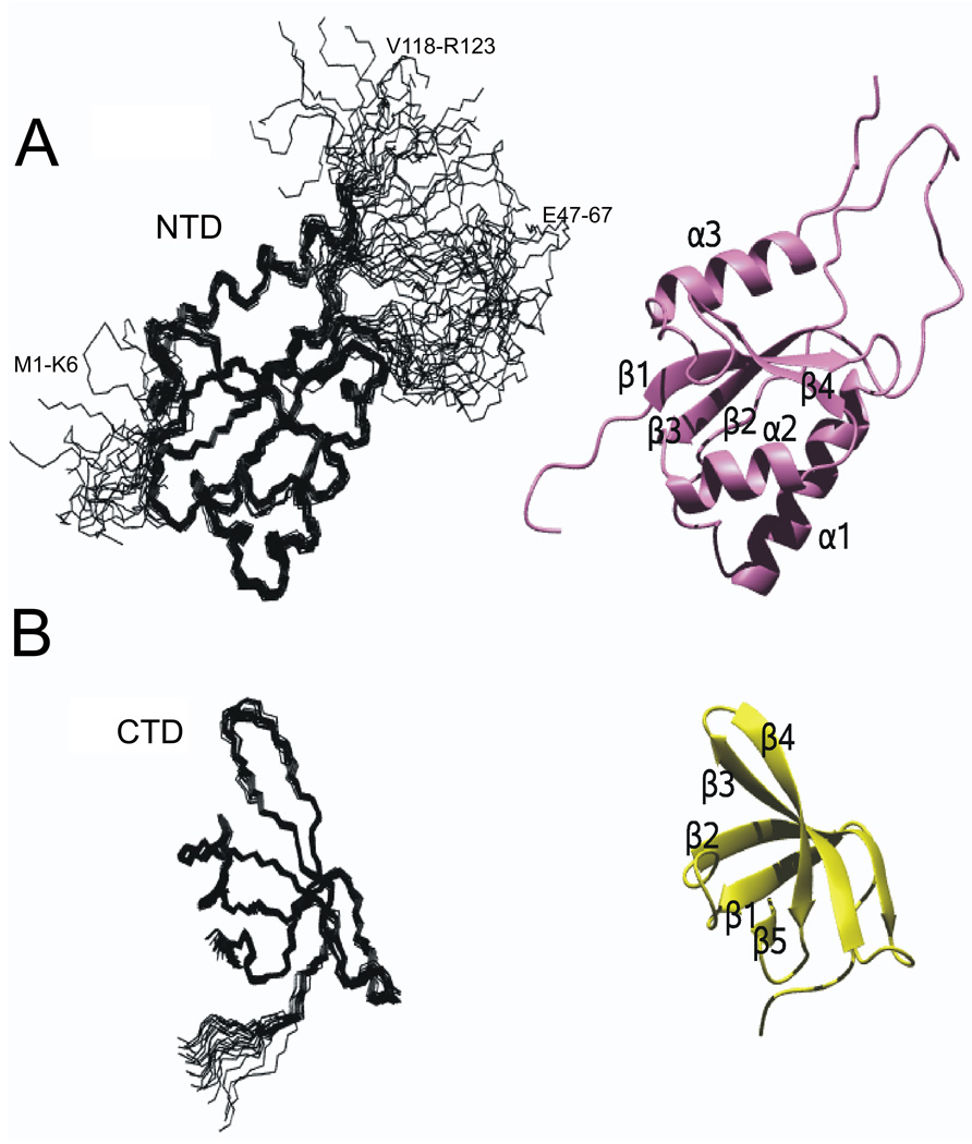
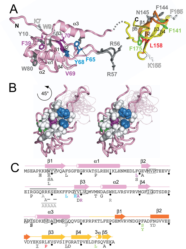
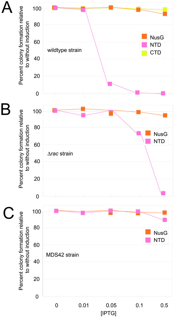
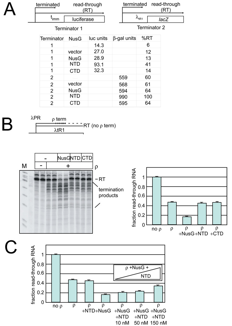
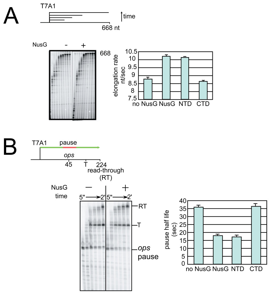
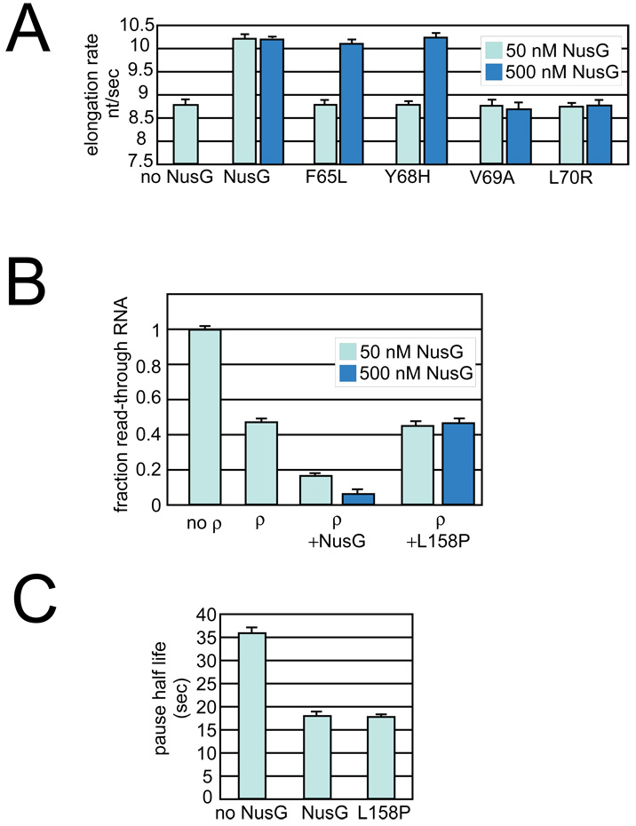
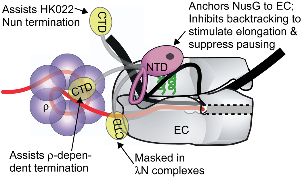
NusG is a conserved regulatory protein that interacts with elongation complexes (ECs) of RNA polymerase, DNA, and RNA to modulate transcription in multiple and sometimes opposite ways. In Escherichia coli, NusG suppresses pausing and increases elongation rate, enhances termination by E. coli rho and phage HK022 Nun protein, and promotes antitermination by lambdaN and in ribosomal RNA operons. We report NMR studies that suggest that E. coli NusG consists of two largely independent N- and C-terminal structural domains, NTD and CTD, respectively. Based on tests of the functions of the NTD and CTD and variants of NusG in vivo and in vitro, we find that NTD alone is sufficient to suppress pausing and enhance transcript elongation in vitro. However, neither domain alone can enhance rho-dependent termination or support antitermination, indicating that interactions of both domains with ECs are required for these processes. We propose that the two domains of NusG mediate distinct interactions with ECs: the NTD interacts with RNA polymerase and the CTD interacts with rho and other regulators, providing NusG with different combinations of interactions to effect different regulatory outcomes.


Figures

Figure 1
Figure 1. Solution structure of E. coli NusG NTD and CTD
(A) Solution structure of EcNusG NTD (left) Structural ensemble of 20 accepted structures; regions of disorder are labeled by residue numbers (M1-K6, P45-F65, V1180-R123). (right) Ribbon representation of representative structure. The structure was determined with experimental NMR restraints obtained from the NTD construct. (B) Solution structure of NusG CTD. (left) Structural ensemble of 20 accepted structures. (right) Ribbon representation of representative structure. The structure was determined with NMR restraints obtained from experiments with FL NusG.
Figure 2
Figure 2. Structure of E. coli NusG
(A) Structures of E. coli NusG. NusG has two main structural domains separated by a linker. The NTD (pink) consists entirely of the NusG N-terminal (NGN) motif, identified only in NusG and its homologs. The CTD (yellow and orange) contains the KOW sequence motif (orange), which has been found in NusG and homologs as well as in ribosomal proteins L24, L26, L27. Mutations characterized in this paper and in previous work (Table 3) are mapped onto the structure and color-coded as follows: green residues fail to complement ΔnusG in vivo but function in vitro, magenta residues are defective for both elongation and termination, blue residues work only at high concentration, the red residue is defective for termination but not elongation, white (outlined) residues exhibited overexpression toxicity but allowed complementation of ΔnusG in vivo and functioned in in vitro assays, and gray residues were previously characterized . Gray dashed line indicates the linker between the two domains and is not intended to suggest its actual position. (B) (left and center) Stereo view showing mutations on one face of the NTD that are part of a hydrophobic pocket, coloring as in (A); (C) Amino acid sequence of EcNusG shown with corresponding structure (above) and substitutions (below). Substitutions are colored as in (A).
Figure 3
Figure 3. Plating efficiency of cells expressing FL NusG, NTD, or CTD
(A) Wild-type MG1655 was tested for growth with plasmids encoding FL NusG, NTD, or CTD. Cells were plated onto media containing different amount of IPTG to induce protein expression. The number of colonies was determined and expressed as a fraction of the number of colonies formed without IPTG (without protein induction). The vertical bars represent the propagated errors from these calculations. (B) Plating efficiency of the Δrac strain (RSW472, Table 1) with plasmids encoding FL NusG or NTD, as described above. (C) Plating efficiency of the MDS42 strain with plasmids encoding FL NusG or NTD, as described above.
Figure 4
Figure 4. In vivo and in vitro ρ-termination
(A) In vivo ρ-termination (Top) In vivo ρ-termination was measured on two terminators, timm (terminator 1) and λtR1 (terminator 2) with downstream reporters. (Bottom) Reporter expression for strains containing plasmids expressing NusG, NTD, and CTD, vector alone, or no plasmid. The %RT is determined by comparison of the levels of expression compared to the levels when ρ is inhibited by bicyclomycin. Luciferase units (RLU) are given as × 106 values. β-galactosidase values are Miller units . (B) In vitro ρ-termination (Top) Schematic of termination template. (Bottom) In vitro transcription reactions were performed in the presence and absence of ρ, FL NusG, NTD, or CTD as indicated. The read-through RNA product is indicated at the top of the gel (labeled RT). Addition of ρ stimulates termination, as seen by a decrease in the amount of the RT product and an increase in the amounts of shorter products (labeled termination products). NusG additionally increases ρ -termination at earlier template positions. (Right). Quantitation of ρ–termination assays. The fraction read-through RNA is determined as the amount of the read-through RNA as a fraction of the total RNA (read-through RNA plus the terminated RNAs). Bars represent averages from three experiments; error is the standard deviation of these averages. (C) Termination assays were performed as in (B), but increasing amounts of NTD (10, 50, and 150 nM) were added together with FL NusG (at 10 nM). NTD competes for RNAP binding with FL NusG as evidenced by the decreased NusG stimulation of termination with increasing amounts of competing NTD. Bars represent averages from three experiments; error is the standard deviation of these values.
Figure 5
Figure 5. The effect of FL NusG, NTD, and CTD on elongation enhancement
(A) Elongation rate assay (top) Schematic of transcription template. Transcription was performed using a template containing the strong T7A1 promoter followed by a U-less cassette allowing formation of halted complexes in the presence of ATP, CTP, and 32P-GTP. (left) After halted complex formation, all four NTPs were added to resume transcription and samples were removed at 5”, 10”, 15”, 20”, 30”, 45”, 1’, 1.5’, 2’, and 4’. Stimulation of transcription elongation by NusG is seen by the faster accumulation of the 668nt product (labeled 668). (right) Quantification of elongation rate in nucleotides synthesized per second for reactions performed in the absence of added factor or with FL NusG, NTD, or CTD. Values plotted are the results from at least three independent experiments; error bars indicate the standard deviation of these averaged values. (B) Pause assay (top) Schematic of ops-containing transcription template. (left)Transcription assays were initiated by forming halted complexes as described above. All four NTPs were then added to resume transcription from the halted complexes through the ops pause sequence; samples were removed at 5”, 15”, 30”, 45”, 1’, and 2’. The effect of NusG is seen by the faster decrease in RNAs paused at the ops pause (labeled ops pause) as ECs escape the pause and the faster accumulation of the longer RNA products (labeled T for products terminating at the downstream terminator or RT for the read-through products). (right) Quantification of pause half-life determined from reactions performed in the absence of added factor, FL NusG, NTD, or CTD. Pause half-life is the amount of time it takes half of the complexes to escape the pause. Values plotted are the results from at least three independent experiments; error bars indicate the standard deviation of these averaged values. Pause efficiencies did not vary significantly with experimental conditions (80–90%).
Figure 6
Figure 6. The effect of NusG mutants on elongation and termination
(A) Mutants near the hydrophobic binding site were tested at higher concentrations for elongation rate enhancement (experiment as shown in Figure 5AFigure 5A). Light blue, 50 nM NusG; Dark blue, 500 nM NusG. (B) The L158P mutation was tested at higher concentrations for enhancement of ρ–termination (experiment as shown in Figure 4BFigure 4B). Light blue, 50 nM NusG; Dark blue, 500 nM NusG. (C) The L158P mutation is able to enhance elongation at a rate comparable to that of wildtype NusG.
Figure 7
Figure 7. Model of NusG interaction
Model of NusG interactions with EC. NusG NTD (pink) is shown interacting with the β’ clamp helices (green) near the upstream edge of the transcription bubble. Alternative interactions of NusG CTD (yellow), for example with ρ, generate NusG’s regulatory complexity. The location of the flexible loop in the NTD and the precise orientation of the domains remain to be determined.

Similar articles

Cited by

References

    1. Burova E, Gottesman ME. NusG overexpression inhibits Rho-dependent termination in Escherichia coli. Mol Microbiol. 1995;17:633–641. - PubMed
    1. Burns CM, Richardson JP. NusG is required to overcome a kinetic limitation to Rho function at an intragenic terminator. Proc Natl Acad Sci U S A. 1995;92:4738–4742. - PMC - PubMed
    1. Artsimovitch I, Landick R. Pausing by bacterial RNA polymerase is mediated by mechanistically distinct classes of signals. Proc Natl Acad Sci U S A. 2000;97:7090–7095. - PMC - PubMed
    1. Pasman Z, von Hippel PH. Regulation of rho-dependent transcription termination by NusG is specific to the Escherichia coli elongation complex. Biochemistry. 2000;39:5573–5585. - PubMed
    1. Yakhnin AV, Yakhnin H, Babitzke P. Function of the Bacillus subtilis transcription elongation factor NusG in hairpin-dependent RNA polymerase pausing in the trp leader. Proc Natl Acad Sci U S A. 2008;105:16131–16136. - PMC - PubMed

Publication types

MeSH terms

LinkOut - more resources