. 2009 Sep;37(9):1084-95.
doi: 10.1016/j.exphem.2009.05.012. Epub 2009 Jun 6.

Cord blood stem cell expansion is permissive to epigenetic regulation and environmental cues

Affiliations

Cord blood stem cell expansion is permissive to epigenetic regulation and environmental cues

Hiroto Araki et al. Exp Hematol. 2009 Sep.

Abstract

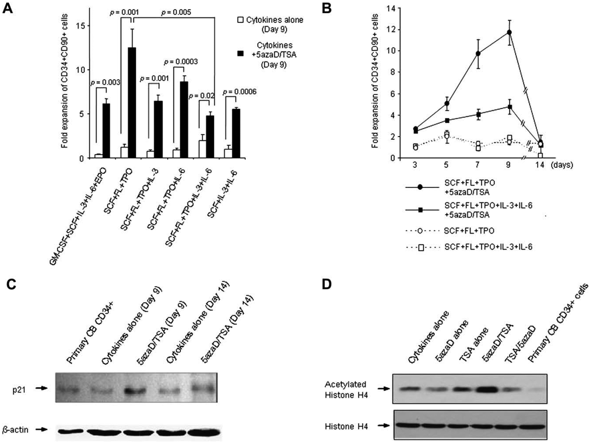
Objective: Augmentation of the number of cord blood (CB) hematopoietic stem cells (HSC) present in a unit is required before it can be considered as an alternative graft for hematopoietic reconstitution for adult patients. In order to further optimize strategies to augment HSC numbers, we examined whether expansion of HSC mediated by epigenetic mechanisms remains permissive to external environmental cues.

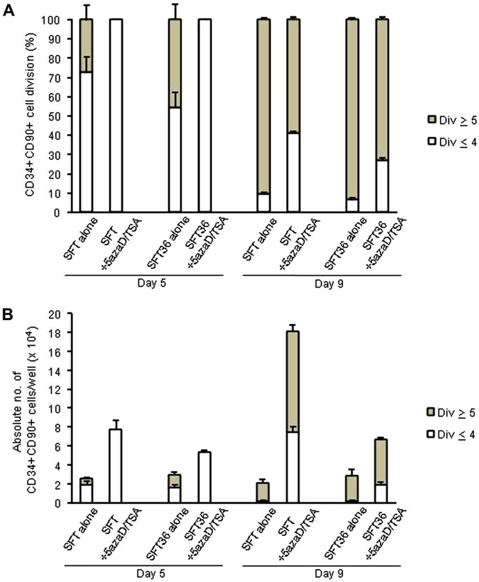
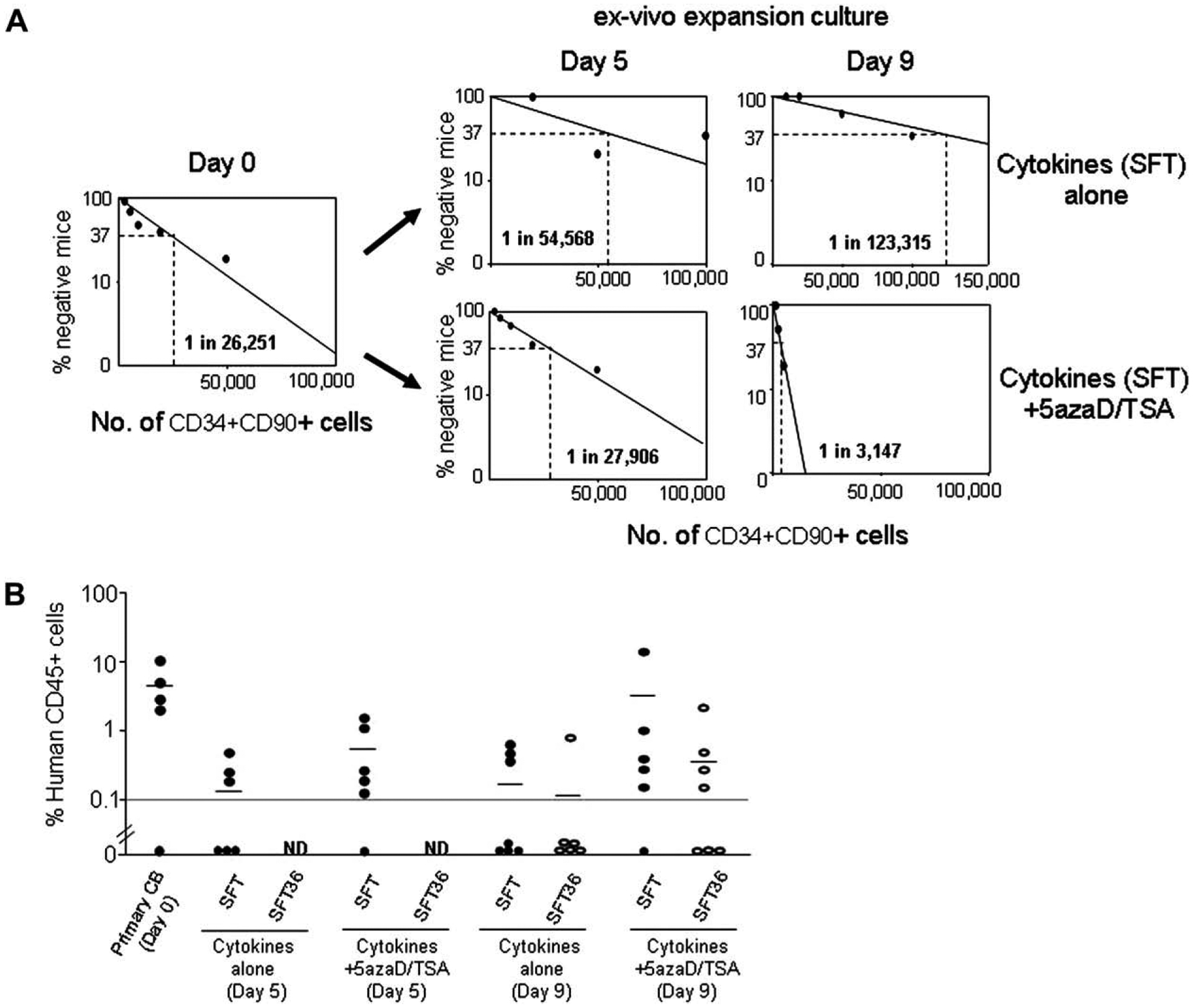
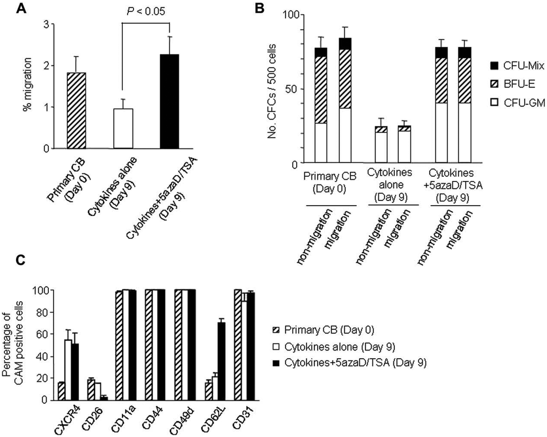
Materials and methods: The chromatin-modifying agents 5-aza-2'-deoxycytidine (5azaD) and trichostatin A (TSA) were used to ameliorate epigenetic alteration of CB cells during ex vivo culture by adding various cytokines. After culture, CD34(+)CD90(+) cell numbers, their division history, in vitro clonogenic potential, and in vivo hematopoietic reconstitution potential and frequency were determined.

Results: 5azaD/TSA-treated, CD34(+)CD90(+) cells were greatly influenced in terms of their degree of expansion, clonogenic potential, cell-division rate, and transplantability by the combination of cytokines used in culture. Furthermore, our current results verify that the sequential addition of 5azaD followed by TSA is crucial for expansion of HSC. We demonstrate that following 5azaD/TSA treatment, the rate of CD34(+)CD90(+) cell division is also dependent on the cytokine cocktail and that this is associated with functional changes, including alteration of in vitro clonogenic potential and in vivo reconstitution potential.

Conclusions: Our studies indicate there are interactions between intrinsic factors influenced by epigenetic mechanisms and external environmental signals in the regulation of HSC expansion. Epigenetic influences on HSC can be accentuated by environmental factors. Regulation of the rate of divisions may be a critical determinant for the maintenance of HSC functional potency during ex vivo expansion.


Conflict of interest statement

Financial disclosure

No financial interest/relationships with financial interest relating to the topic of this article have been declared.

Figures

Figure 1.
Figure 1.
Effects of different cytokine combinations on the expansion of cord blood (CB) CD34+CD90+ cells. (A) Effect of 5-aza-2′-deoxycytidine (5azaD)/trichostatin A (TSA) on the number of CB CD34+CD90+ cells following 9 days of culture in the presence of various cytokine combinations. Cytokine combinations tested during the final 7 days of the 9 day culture were as follows: granulocyte-macrophage colony-stimulating factor (GM-CSF), stem cell factor (SCF), interleukin (IL)-3, IL-6, erythropoietin (EPO); SCF, FL, TPO (SFT); SCF, FL, TPO, IL-3; SCF, FL, TPO, IL-6; SCF, FL, TPO, IL-3, IL-6 (SFT36); SCF, IL-3, IL-6. CD34+ cells (5 × 10⁴/well) were cultured in the presence of SCF, FL, TPO, and IL-3 for the first 48 hours and then the cocktail of cytokines was changed to the designated combinations. The graph represents mean ± standard error of three independent experiments. (B) Effect of 5azaD/TSA on the number of CD34+CD90+ cells following ex vivo culture in various cytokine combinations. The fold expansion was determined at various time points by dividing the output number of CB CD34+CD90+ cells by the input number of CD34+CD90+ cells. (C) Protein levels of P21 were examined on CB cells at day 0, and on cultured CB cells with cytokines (SFT) alone or cytokines (SFT) and 5azaD/TSA at day 9 and day 14 by Western blot analysis. Equal loading of protein was verified with anti – β-actin antibody on the same membrane. (D) Status of histone H4 acetylation of CD34+ cells was examined by Western blot analysis to test the effects of the alteration of the sequence of addition of 5azaD and TSA. Parallel cultures were set up with CB CD34+ cells with cytokines (SFT) alone and SFT with the addition of chromatin-modifying agents; 5azaD alone, TSA alone, TSA followed by 5azaD and 5azaD followed by TSA for 72 hours. Cells were harvested and equal amounts of proteins were electrophoresed on 15% sodium dodecyl sulfate polyacrylamide gel electrophoresis gel. Acetylated-histone H4 level was checked by using anti-acetylated H4 antibody. Histone H4 level was used as a loading control.
Figure 2.
Figure 2.
Effects of 5-aza-2′-deoxycytidine (5azaD)/trichostatin A (TSA) treatment and cytokine combinations on cell division history of CD34+CD90+ cells after 5 and 9 days of culture. (A) Effects of 5azaD/TSA treatment and cytokine combinations on the cell division history of CD34+CD90+ cells. (B) Absolute number of CD34+CD90+ cells generated from CD34+CD90+ cells undergoing five or fewer divisions or five or more divisions present in an individual well were determined at day 5 and at day 9. Primary CB CD34+ cells were labeled with carboxyfluorescein diacetate succinimidyl ester (CFSE) on day 0. After 5 and 9 days of culture, cells were labeled with anti–CD34-allophycocyanin and anti–CD90-phycoerythrin and CD34+CD90+ gated cells were analyzed for a progressive decline of CFSE fluorescence intensity. Data represents the mean ± standard error of three independent experiments.
Figure 3.
Figure 3.
Marrow repopulating potential of the CD34+CD90+ cell population following ex vivo culture. (A) Frequency of SCID mouse repopulating cells (SRC) present in the primary CD34+CD90+ cells prior to (day 0) and following culture in the presence or absence of 5-aza-2′-deoxycytidine (5azaD)/trichostatin A (TSA) treatment (day 5 and day 9) were determined by limiting dilution analysis following transplantation of cord blood (CB) cells in a NOD/SCID mouse assay. Increasing numbers (1,000; 2,000; 5,000; 10,000; 20,000; 50,000; 100,000) of primary CB CD34+CD90+ cells or the progeny of the same number of input CD34+CD90+ cells after 5 and 9 days of ex vivo culture in the presence of SFT were transplanted into NOD/SCID mice. NOD/SCID mice were transplanted with primary CB CD34+ cell fraction or the cellular products of culture lacking 5azaD/TSA treatment (cytokines alone, SFT) or 5azaD/TSA-treated cultures containing cytokines (SFT) initiated with identical numbers of CD34+CD90+ cells. The frequency of SRC in primary CB CD34+CD90+ cells was 1 in 26,251 (95% confidence interval: 1/10,627 – 1/64,850), 1 in 54,568 (95% confidence interval: 1/16,909 − 1/176,101) in the culture containing cytokines alone (day 5) and 1 in 123,315 (95% confidence interval: 1/46,617 – 1/326,200) in the culture containing cytokines alone (day 9). The frequency of SRC was 1 in 27,906 (95% confidence interval: 1/10,280 – 1/75,748) in the 5azaD/TSA-treated cells cultured for 5 days and 1 in 3,147 (95% confidence interval: 1/1,602 – 1/6,189) in the 5azaD/TSA-treated cells cultured for 9 days. Data was analyzed by applying Poisson statistics according to the single-hit model. (B) NOD/SCID engraftment observed with the transplantation of expanded cells containing 5 × 10⁴ of CD34+CD90+ cells per mouse following 5 and 9 days of culture in the presence or absence of 5azaD/TSA with SCF + FL + TPO (SFT) (●) or SCF + FL + TPO + IL- 3 + IL-6 (SFT36) (○).
Figure 4.
Figure 4.
Migration and levels of cell adhesion molecules expressed in primary cord blood (CB) CD34+ cells cultured with or without 5-aza-2′-deoxycytidine (5azaD)/trichostatin A (TSA). (A) Migration of cells through a preformed basement membrane (Matrigel). (B) Plating efficiency of the cells that migrated through Matrigel compared with that of nonmigrating cells. Equal numbers of cells (500 cells) from the upper (nonmigrating) and lower (migrating) compartments of the Boyden chamber were plated and colonies were scored after 14 days of incubation. (C) Primary CD34+CD90+ cells and CD34+CD90+ cells cultured with or without 5azaD/TSA were analyzed for cell adhesion molecules and CXCR4 expression profiles. Cytokine cocktail used for these experiments included interleukin-3 (IL-3), stem cell factor (SCF), Flt3 ligand (FL), and thrombopoietin (TPO) for the initial 48 hours, while for the remaining 7 days of culture the cytokine cocktail changed to SCF, FL, and TPO. After 9 days of ex vivo culture, CD34+CD90+ cells were examined for cell adhesion molecules and CXCR4 expression using three-color fluorescence analysis. Each value represents mean ± standard error of three independent experiments.

References

    1. Blau HM. Differentiation requires continuous active control. Annu Dev Biochem. 1992;61:1213–1230. - PubMed
    1. Jones PA, Takai D. The role of DNA methylation in mammalian epigenetics. Science. 2001;293:1068–1070. - PubMed
    1. Marks PA, Richon VM, Rifkind RA. Histone deacetylases inhibitors: inducers of differentiation or apoptosis of transformed cells. J Natl Cancer Inst. 2000;92:1210–1216. - PubMed
    1. Kass SU, Pruss D, Wolffe AP. How dose methylation mediated repress transcription? Trends Genet. 1997;13:444–449. - PubMed
    1. Issa JP. Decitabine. Curr Opin Oncol. 2003;15:446–451. - PubMed

Publication types

MeSH terms