Comparative Study
. 2009 Sep;37(9):1038-53.
doi: 10.1016/j.exphem.2009.05.011. Epub 2009 Jun 6.

VEGF and IHH rescue definitive hematopoiesis in Gata-4 and Gata-6-deficient murine embryoid bodies

Affiliations
Comparative Study

VEGF and IHH rescue definitive hematopoiesis in Gata-4 and Gata-6-deficient murine embryoid bodies

Monique Pierre et al. Exp Hematol. 2009 Sep.

Abstract

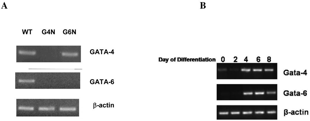
Objective: Murine embryonic stem cells can be differentiated into embryoid bodies (EBs), which serve as an in vitro model recapitulating many aspects of embryonic yolk sac hematopoiesis. Differentiation of embryonic stem cells deficient in either Gata-4 or Gata-6 results in EBs with disrupted visceral endoderm (VE). While lack of VE has detrimental effects on hematopoiesis in vivo, it is unclear whether lack of VE affects hematopoiesis in EBs. Therefore, we compared Gata-4 null (G4N) and Gata-6 null (G6N) EBs with wild-type EBs to assess their ability to commit to hematopoietic cells.

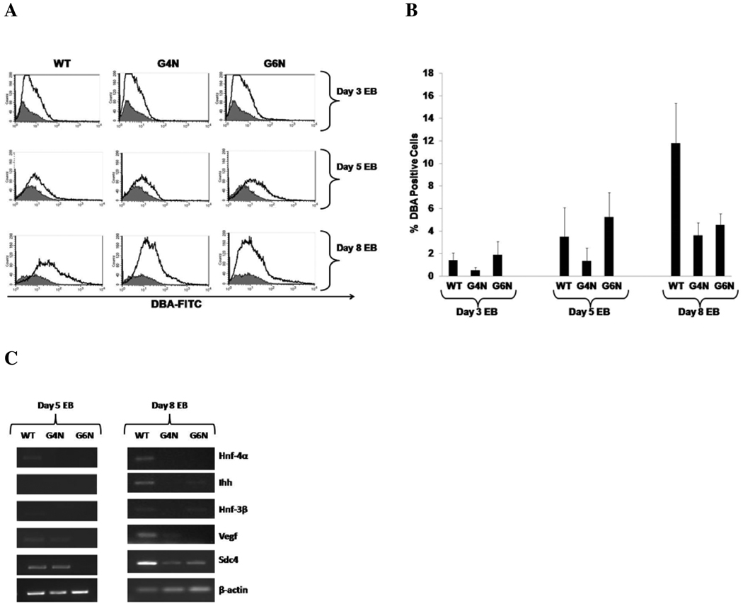
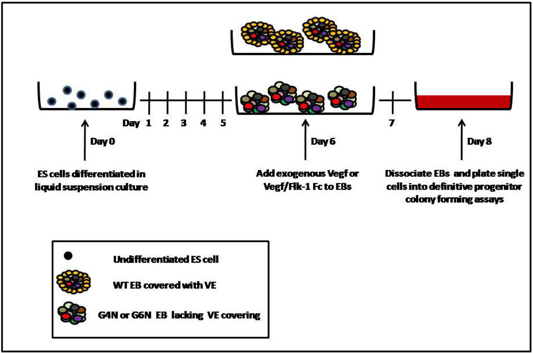
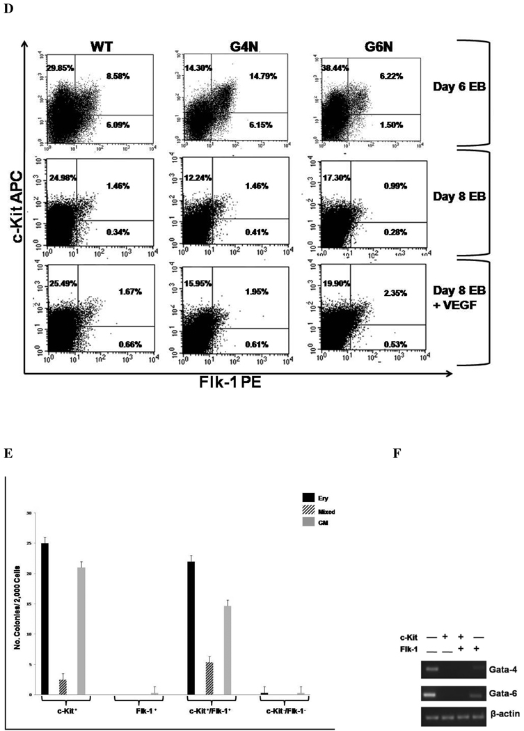
Materials and methods: EB VE formation was examined using cell-sorting techniques and analysis visceral endoderm gene expression. Hematopoietic progenitor potential of EBs cultured under various conditions was assessed using colony-forming assays.

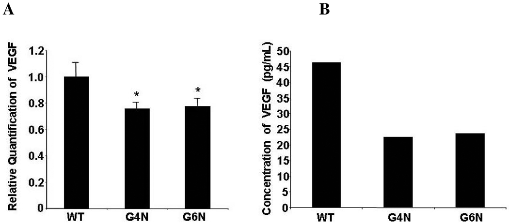
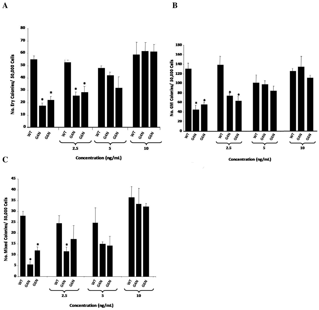
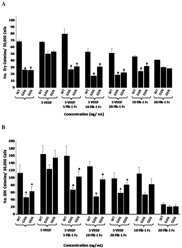
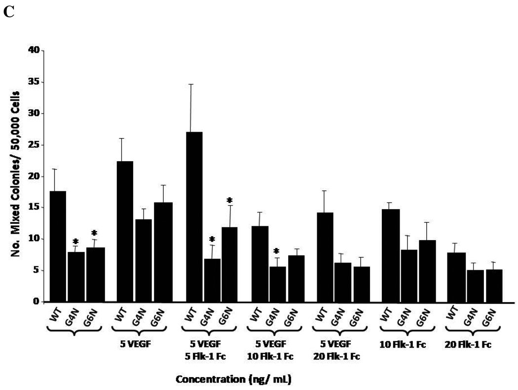
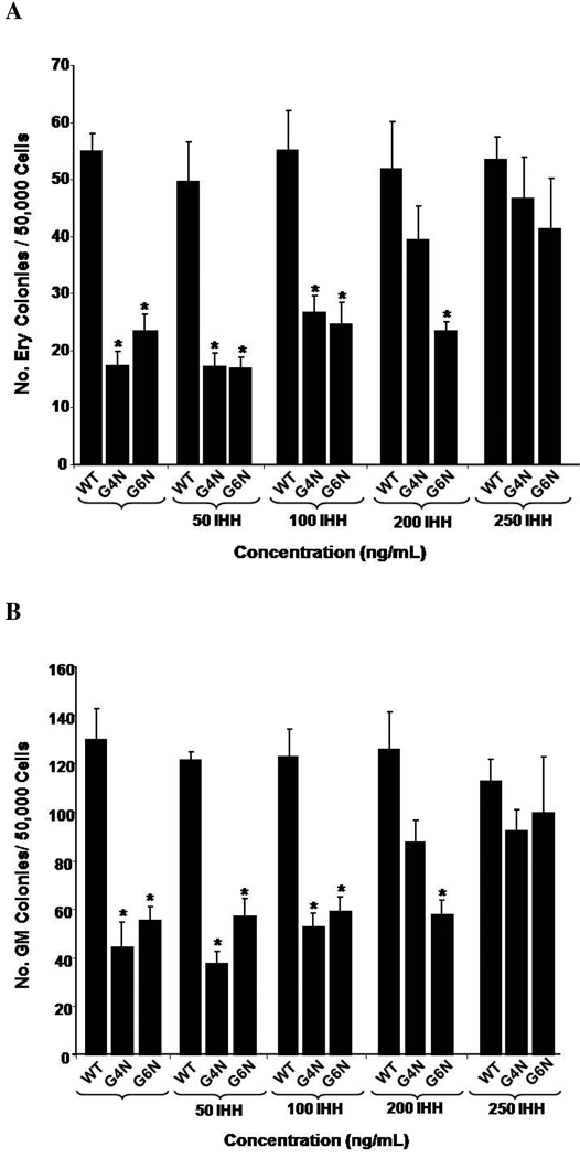
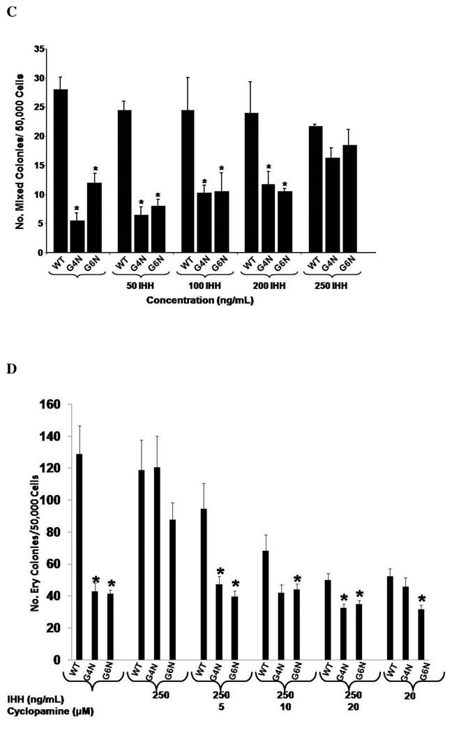
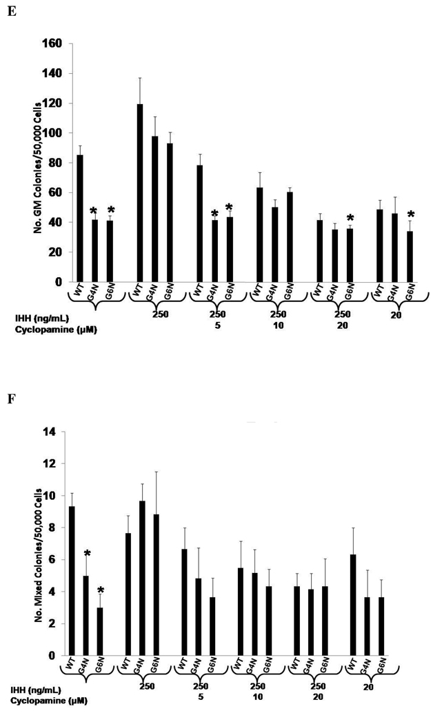
Results: Definitive erythroid, granulocyte-macrophage, and mixed colonies were significantly reduced in G4N and G6N EBs compared to wild-type EBs. Vascular endothelial growth factor (VEGF) expression and secretion were also reduced in both G4N and G6N EBs, consistent with VE serving as a site of VEGF production. Addition of exogenous VEGF(165), to EB cultures completely rescued definitive colony-forming cells in G4N and G6N EBs. This rescue response could be blocked by addition of soluble Flk-1 Fc to EB cultures. Similarly, addition of exogenous Indian hedgehog to EB cultures also recovers the diminishment in definitive hematopoiesis in a reversible manner.

Conclusion: These results suggest that the absence of VE in G4N and G6N EBs does not prevent emergence of definitive progenitors from EBs. However, the decreased level of VEGF and Indian hedgehog production in VE devoid G4N and G6N EBs attenuates definitive hematopoietic progenitor cell expansion.


Figures

Figure 1
Figure 1. GATA-4 and GATA-6 transcripts were detected in WT EBs
Total cellular RNA was isolated from WT, G4N and G6N day 8 EBs (A) and day 0, 2, 4, 6 and 8 WT EBs (B). Reverse transcription was performed using 0.8 µg as template. GATA-4, GATA-6 and β-actin were amplified with 28 cycles of PCR. Products were visualized on agarose gel stained with ethidium bromide.
Figure 2
Figure 2. Diminished Visceral Endoderm Formation and Gene Expression in G4N and G6N EBs
Cells from day 3, day 5 and day 6 EBs were stained with FITC-conjugated DBA and positive bound cells were analyzed by FACS analysis. (Representative experiment) (A). The average of four independent experiments are displayed (B). DBA positively bound cells were sorted from day 5 and day8 WT, G4N and G6N EBs. Total cellular RNA was isolated and reverse transcription was performed using 2.0 µg as template. Hnf-4α, Ihh, Hnf-3β, Vegf, Sdc4 and β-actin were amplified with 28 cycles of PCR. Products were visualized on agarose gel stained with ethidium bromide (C).
Figure 3
Figure 3. Definitive hematopoiesis is diminished in G4N and G6N day 8 EBs
Cells from day 8, 9 and 10 EBs were plated as single cells into definitive progenitor assays, and definitive erythroid (Ery), granulocyte-macrophage (GM) and mixed (Mixed) lineage colonies were scored after 7 days. (N = 5 independent experiments). Error bars represent s.e.m. *p<0.05.
Figure 4
Figure 4. VEGF mRNA levels and protein secretion are reduced in G4N and G6N EBs
Total RNA was isolated from day 6 EBs. RNA was reverse transcribed and real-time PCR analysis was performed using the ABI 7500 for Vegf relative quantification. Error bars represent s.e.m. *p<0.05 (A). Supernatant from day 8 EBs was collected, and the concentration of VEGF protein was determined by ELISA (representative experiment) (B).
Figure 5
Figure 5. Schematic diagram of VEGF and Flk-1 Fc treatment of EB cultures
In rescue experiments, EBs were treated with VEGF (2.5, 5 and 10 ng/mL) on EB differentiation day 6 and allowed to differentiate for 2 additional days. In blocking experiments, 5 ng/mL of VEGF and soluble Flk-1 Fc (5, 10, 20 ng/mL) were added to the culture medium on EB differentiation day 6 and EBs were differentiated for 2 additional days. In both cases, EBs were dissociated into single cells on day 8 and plated into definitive hematopoietic progenitor assays.
Figure 6
Figure 6. VEGF rescues definitive hematopoiesis in G4N and G6N EBs
EBs were cultured for 6 days and VEGF (2.5, 5, 10 ng/ mL) was added to the culture medium. After 2 additional days, day 8 EBs were dissociated into single cells and plated into definitive hematopoietic progenitor colony formation assays. Definitive Erythroid (A), GM (B), and mixed (C) colonies were scored after 7 days. (N= 3 independent experiments). Error bars represent s.e.m. *p<0.05. EBs were dissociated into single cells on day 6 or on day 8 after being either treated or untreated with Vegf (5 ng/mL) on day 6. Cells were then stained with PE-conjugated c-Kit and APC-conjugated antibodies and analyzed by FACS analysis (representative experiment of 3 independent experiments) (D). Sorted cell populations were plated into definitive hematopoietic progenitor colony formation assays (representative experiment) (E.) Total cellular RNA was isolated from sorted cell populations and reverse transcription was performed using 0.8 µg as template. Gata-4 and Gata-6 expression were analyzed by RT-PCR (F). β-actin served as the positive control. Products were amplified with 28 cycles of PCR and visualized on agarose gel stained with ethidium bromide.
Figure 7
Figure 7. VEGF mediated rescue of definitive hematopoiesis in day 8 EBs is abolished with soluble Flk-1/Fc
EBs were cultured for 6 days and 5 ng/mL of VEGF and soluble Flk-1/ Fc (5, 10, 20 ng/mL) were added to the culture medium. After 2 additional days, day 8 EBs were dissociated into single cells and plated into definitive hematopoietic progenitor colony formation assays. Definitive Erythroid (A), GM (B), and mixed (C) colonies were scored after 7 days. (N= 3 independent experiments). Error bars represent s.e.m. *p<0.05.
Figure 8
Figure 8. IHH specifically rescues definitive hematopoiesis in G4N and G6N EBs
EBs were cultured for 6 days and IHH (50, 100, 200, 250 ng/ mL) was added to the culture medium. After 2 additional days, day 8 EBs were dissociated into single cells and plated into definitive hematopoietic progenitor colony formation assays. Definitive Erythroid (A), GM (B), and mixed (C) colonies were scored after 7 days. EBs were cultured for 6 days and IHH (250 ng/ml) and cylopamine (5, 10, 20 µM) were added and EB cells were plated after two additional days of culture into definitive hematopoietic progenitor colony forming assays. Colonies were scored as definitive erythroid (D), GM (E), and mixed (F) after 7 days. (N= 3 independent experiments). Error bars represent s.e.m. *p<0.05

References

    1. Moore MA, Metcalf D. Ontogeny of the haemopoietic system: yolk sac origin of in vivo and in vitro colony forming cells in the developing mouse embryo. Br J Haematol. 1970;18:279–296. - PubMed
    1. Palis J, Robertson S, Kennedy M, Wall C, Keller G. Development of erythroid and myeloid progenitors in the yolk sac and embryo proper of the mouse. Development. 1999;126:5073–5084. - PubMed
    1. Lux CT, Yoshimoto M, McGrath K, Conway SJ, Palis J, Yoder MC. All primitive and definitive hematopoietic progenitor cells emerging before E10 in the mouse embryo are products of the yolk sac. Blood. 2008;111:3435–3438. - PMC - PubMed
    1. Sabin F. Studies on the origin of blood vessels and of red blood corpuscules as seen in the living blastoderm of chicks during the second day of incubation. Contributions to Embryology. Carnegie Inst. Pub. 1920:214–262.
    1. Murray P. The develpment in vitro of blood of the early chick embryo. Proc Roy Soc. 1932;111:497–521.

Publication types

MeSH terms

Substances