

Accuracy of volumetric bone mineral density measurement in high-resolution peripheral quantitative computed tomography
- PMID: 19501201
- PMCID: PMC4454742
- DOI: 10.1016/j.bone.2009.05.023
Accuracy of volumetric bone mineral density measurement in high-resolution peripheral quantitative computed tomography
Abstract
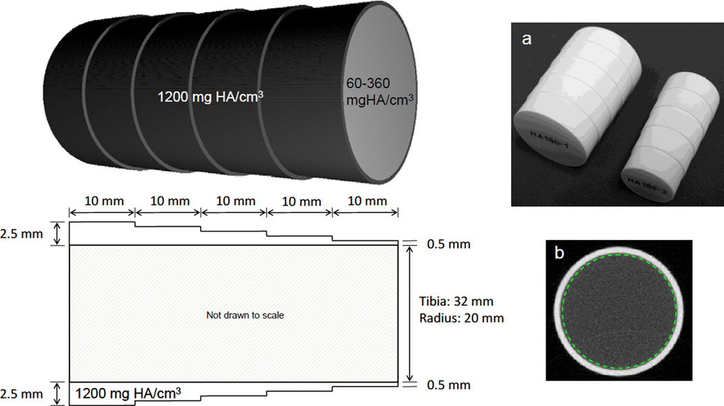
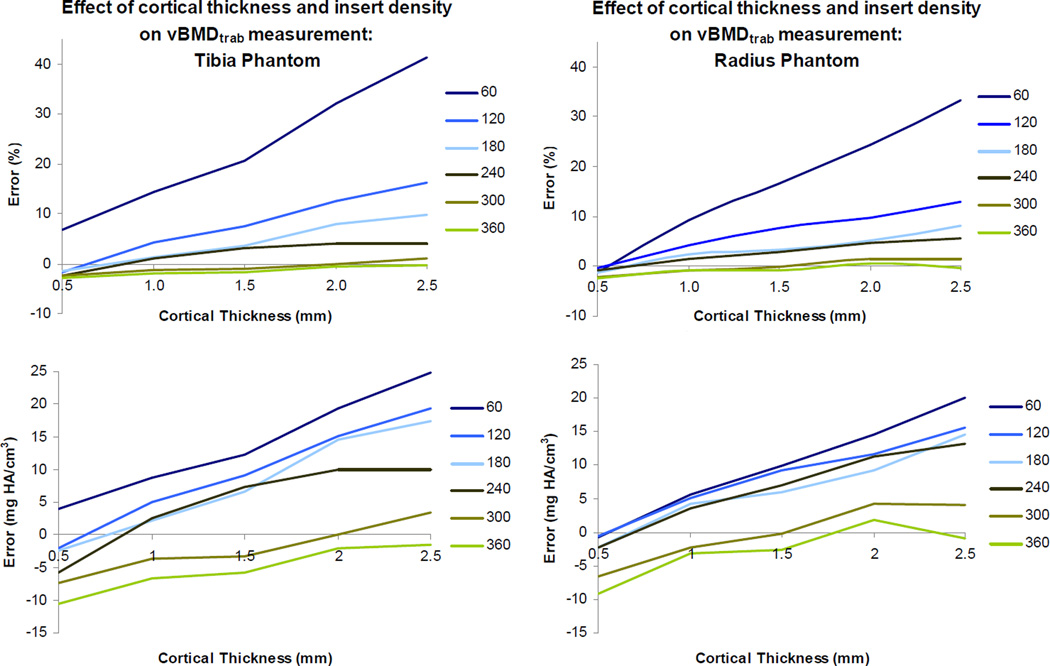
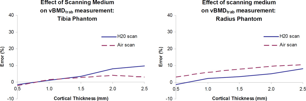
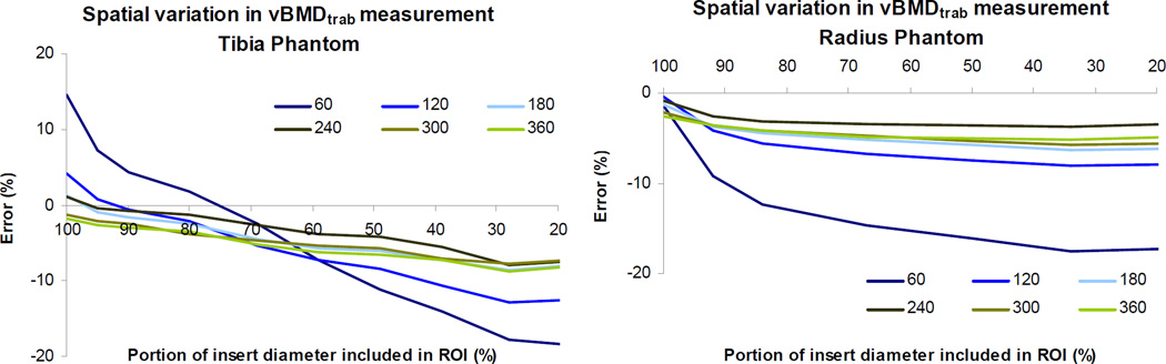
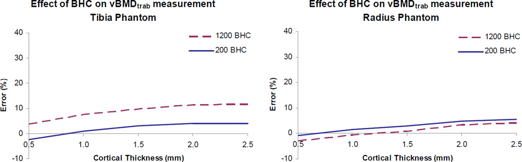
Accurate bone mineral density (BMD) quantification is critical in clinical assessment of fracture risk and in the research of age-, disease-, and treatment-related musculoskeletal changes. The development of high-resolution peripheral quantitative computed tomography (HR-pQCT) imaging has made possible in vivo assessment of compartmental volumetric BMD (vBMD) and bone micro-architecture in the distal radius and tibia. HR-pQCT imaging relies on a polychromatic X-ray source and therefore is subject to beam hardening as well as scatter artifacts. In light of these limitations, we hypothesize that the accuracy of HR-pQCT vBMD measurement in the trabecular compartment (vBMD(trab)) is not independent of bone density and geometry, but rather influenced by variations in trabecular bone volume fraction and cortical thickness. The goal of this study, therefore, was to evaluate the accuracy of HR-pQCT vBMD(trab) measurement in the radius and tibia, and to determine the dependence of this measurement on geometric and densitometric parameters. Our approach was to use a series of idealized hydroxyapatite (HA) phantoms with varying densities and geometries to quantify the accuracy of HR-pQCT analysis. Two sets of custom-made HA phantoms designed to mimic the distal tibia and distal radius were manufactured. Geometric and densitometric specifications were based on a dataset of healthy volunteers and osteopenic patients. Multiple beam hardening correction (BHC) algorithms were implemented and evaluated in their ability to reduce measurement error. Substantial errors in measured vBMD(trab) were found. Overestimation of vBMD(trab) increased proportional to cortical shell thickness and decreased proportional to insert density. The most pronounced vBMD(trab) overestimation therefore occurred in the phantoms with the lowest insert densities and highest shell thickness, where error was as high as 20 mg HA/cm3 (33%) in the radius phantom and 25 mg HA/cm(3) (41%) in the tibia phantom. Error in vBMD(trab) propagates to the calculation of micro-architectural measures; 41% error in vBMD(trab) will produce 41% error in volume fraction (BV/TV) and trabecular thickness (Tb.Th), and 5% error in trabecular separation (Tb.Sp). BHC algorithms supplied by the manufacturer failed to eliminate these errors. Our results confirm that geometric and densitometric variations influence the accuracy of HR-pQCT vBMD(trab) measurements, and must be considered when interpreting data across populations or time-points.
Figures
References
-
- Black DM, Cummings SR, Karpf DB, Cauley JA, Thompson DE, Nevitt MC, Bauer DC, Genant HK, Haskell WL, Marcus R, Ott SM, Torner JC, Quandt SA, Reiss TF, Ensrud KE. Randomised trial of effect of alendronate on risk of fracture in women with existing vertebral fractures. Fracture Intervention Trial Research Group. Lancet. 1996;348:1535–1541. - PubMed
-
- Cummings SR. How drugs decrease fracture risk: Lessons from trials. J Musculoskelet Neuronal Interact. 2002;2:198–200. - PubMed
-
- Beck TJ, Looker AC, Ruff CB, Sievanen H, Wahner HW. Structural trends in the aging femoral neck and proximal shaft: analysis of the Third National Health and Nutrition Examination Survey dualenergy X-ray absorptiometry data. J Bone Miner Res. 2000;15:2297–2304. - PubMed
-
- Sornay-Rendu E, Boutroy S, Munoz F, Delmas PD. Alterations of cortical and trabecular architecture are associated with fractures in postmenopausal women, partially independent of decreased BMD measured by DXA: the OFELY study. J Bone Miner Res. 2007;22:425–433. - PubMed
Publication types
MeSH terms
Grants and funding
LinkOut - more resources
Full Text Sources
Medical
