Review
. 2009 Aug;21(4):233-41.
doi: 10.1016/j.smim.2009.05.004. Epub 2009 Jun 6.

Autophagy as an emerging dimension to adaptive and innate immunity

Affiliations
Review

Autophagy as an emerging dimension to adaptive and innate immunity

Séamus Hussey et al. Semin Immunol. 2009 Aug.

Abstract

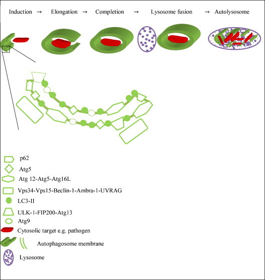
Autophagy is an evolutionary conserved cellular process during which cytoplasmic material is engulfed in double membrane vacuoles that then fuse with lysosomes, ultimately degrading their cargo. Emerging evidence, however, now suggests that autophagy can form part of our innate and adaptive immune defense programs. Recent studies have identified pattern recognition molecules as mediators of this process and shown that intracellular pathogens can interact with and even manipulate autophagy. Recent translational evidence has also implicated autophagy in the pathogenesis of several immune-mediated diseases, including Crohn disease. In this review, we present autophagy in the context of its role as an immune system component and effector and speculate on imminent and future research directions in this field.


Figures

Fig. 1
Fig. 1
Autophagosome biogenesis. The earliest identifiable structure in the initiation (nucleation) sequence of autophagosome formation is the crescent-shaped isolation membrane or phagophore. Key elements include Atg9, the ULK1–FIP200–Atg13 complex, LC3-II, the Atg12–Atg5–Atg16L complex. Once formed, this membrane progressively elongates (elongation), encircling its cytosolic target, e.g. bacterium, within a portion of the cytosol. The membrane tips fuse and eventually seal, forming the autophagosome (completion). The autophagosome may fuse with the endosomal compartment, forming an amphisome, prior to its ultimate maturation step, whereby its outer membrane fuses with the lysosome to form an autolysosome (also termed autophagolysosome). This facilitates degradation, processing and recycling of the contents of the autophagosome.
Fig. 2
Fig. 2
TLR activation triggers autophagy. LPS triggers autophagy after recruitment of Trif (also RIP1 and p38, not shown) and MyD88. The latter seems to interact with Beclin-1, reducing its binding to the anti-autophagic molecule BCL-2. TLR2 engagement induces the incorporation of LC3 to phagosomes (unkown mechanism). Viruses are able to induce autophagy through TLR3, RIG-I (dsRNA) or TLR7/8-MyD88 (ssRNA). Conventional DCs sense viral ligads through the RIG-I/MAVS axis to secrete type I interferon, while the conjugate ATG5/12 seems to be a down regulator of such response. Plasmocytoid DCs deliver TLR7 ligands from the cytosol to the compartments containing TLR7 using basal autophagy. IPAF inhibits autophagy through an unclear mechanism.

References

    1. Klionsky D.J. Autophagy: from phenomenology to molecular understanding in less than a decade. Nat Rev Mol Cell Biol. 2007;8:931–937. - PubMed
    1. Ashford T.P., Porter K.R. Cytoplasmic components in hepatic cell lysosomes. J Cell Biol. 1962;12:198–202. - PMC - PubMed
    1. Clark S.L., Jr. Cellular differentiation in the kidneys of newborn mice studies with the electron microscope. J Biophys Biochem Cytol. 1957;3:349–362. - PMC - PubMed
    1. Hruban Z., Spargo B., Swift H., Wissler R.W., Kleinfeld R.G. Focal cytoplasmic degradation. Am J Pathol. 1963;42:657–683. - PMC - PubMed
    1. Kovacs A.L., Seglen P.O. Inhibition of hepatocytic protein degradation by inducers of autophagosome accumulation. Acta Biol Med Ger. 1982;41:125–130. - PubMed

Publication types