. 2009 Aug;297(2):L299-308.
doi: 10.1152/ajplung.00008.2009. Epub 2009 Jun 5.

FGF signaling is required for myofibroblast differentiation during alveolar regeneration

Affiliations

FGF signaling is required for myofibroblast differentiation during alveolar regeneration

Anne-Karina T Perl et al. Am J Physiol Lung Cell Mol Physiol. 2009 Aug.

Abstract

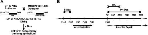
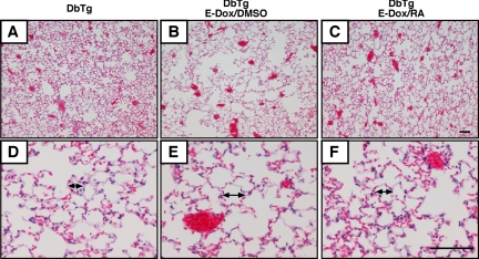
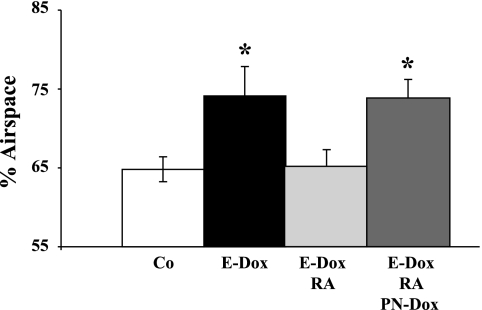
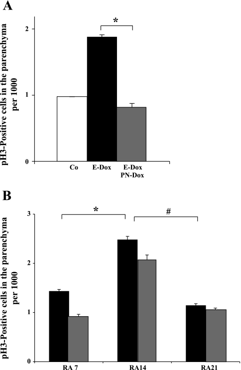
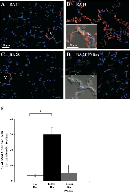
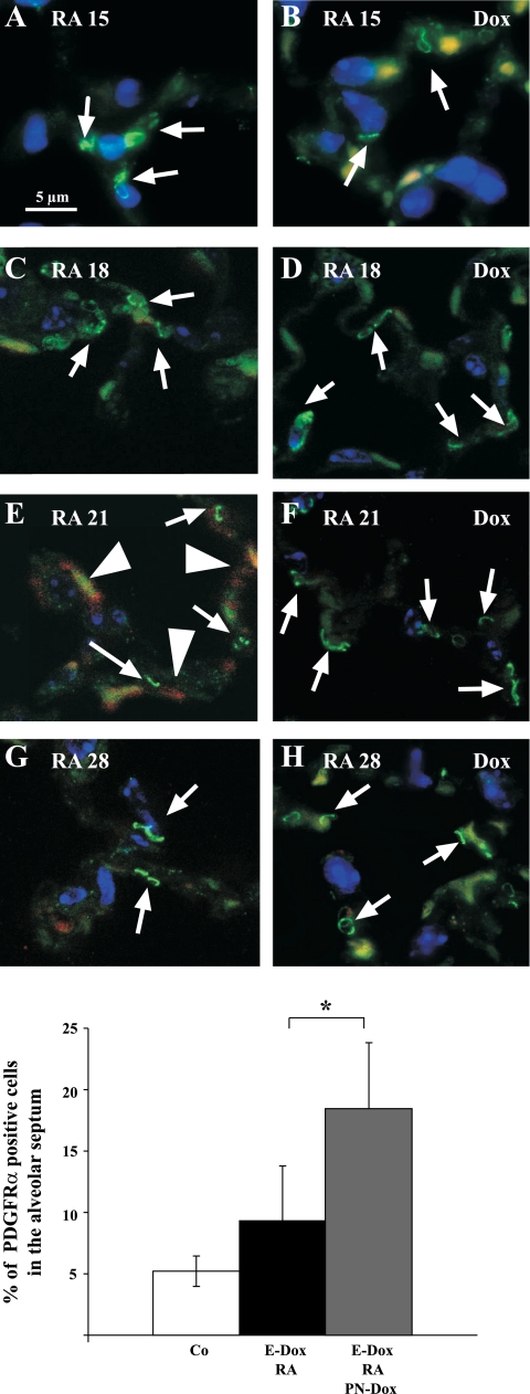
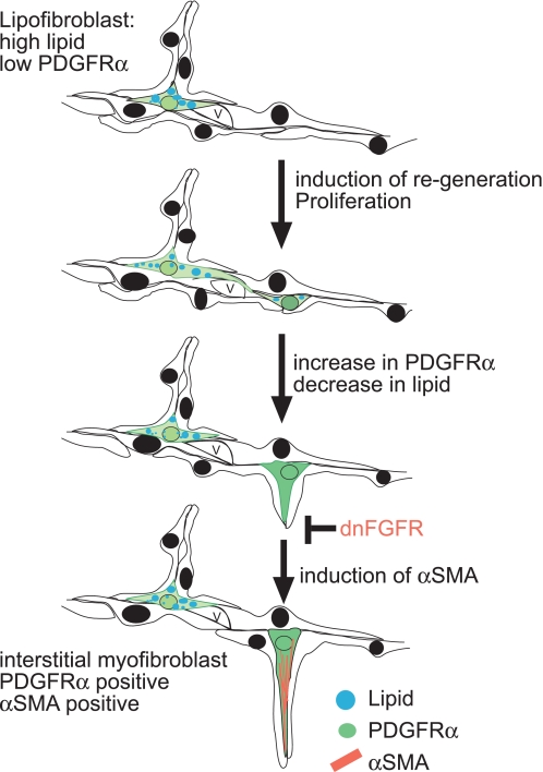
Normal alveolarization has been studied in rodents using detailed morphometric techniques and loss of function approaches for growth factors and their receptors. However, it remains unclear how these growth factors direct the formation of secondary septae. We have previously developed a transgenic mouse model in which expression of a soluble dominant-negative FGF receptor (dnFGFR) in the prenatal period results in reduced alveolar septae formation and subsequent alveolar simplification. Retinoic acid (RA), a biologically active derivative of vitamin A, can induce regeneration of alveoli in adult rodents. In this study, we demonstrate that RA induces alveolar reseptation in this transgenic mouse model and that realveolarization in adult mice is FGF dependent. Proliferation in the lung parenchyma, an essential prerequisite for lung regrowth was enhanced after 14 days of RA treatment and was not influenced by dnFGFR expression. During normal lung development, formation of secondary septae is associated with the transient presence of alpha-smooth muscle actin (alphaSMA)-positive interstitial myofibroblasts. One week after completion of RA treatment, alphaSMA expression was detected in interstitial fibroblasts, supporting the concept that RA-initiated realveolarization recapitulates aspects of septation that occur during normal lung development. Expression of dnFGFR blocked realveolarization with increased PDGF receptor-alpha (PDGFRalpha)-positive cells and decreased alphaSMA-positive cells. Taken together, our data demonstrate that FGF signaling is required for the induction of alphaSMA in the PDGFRalpha-positive myofibroblast progenitor and the progression of alveolar regeneration.


Figures

Fig. 1.
Fig. 1.
A: schematic drawing of the mouse breeding to generate double transgenic (DbTg) mice. Heterocygote mice of the activator line, SP-C-rtTA line 1, are crossed to heterocygote mice of the operator line, tetOdnFGFR-Hfc. According to Mendelian inheritance, only 25% of the offspring carry both transgenes, DbTg mice. Only in DbTg progenies doxycycline (dox) treatment activates dominant-negative FGF receptor (dnFGFR) expression specifically in the lung epithelium (20). B: timeline of dox and retinoic acid (RA) treatment to induce alveolar simplification and alveolar repair, respectively, in DbTg mice. Dox treatment from embryonic day 14.5 (E14.5) to E18.5 (E-Dox) induces expression of dnFGFR and inhibits postnatal alveolarization. RA treatment from postnatal day 35 (PN35) to PN48 induces alveolar repair. Postnatal dox treatment from PN35 throughout RA treatment (PN-Dox) induces dnFGFR expression and inhibits alveolar repair. Alveolar repair in the presence and absence of FGF signaling was assessed in DbTg mice after embryonic inhibition of FGF signaling 7, 14, 21, 28, and 35 days after initiation of RA treatment (RA7, RA14, RA21, RA28, and RA35). SV40, simian virus 40.
Fig. 2.
Fig. 2.
RA improves alveolar simplification in DbTg mice. Lung histology was analyzed by hematoxylin-eosin (H&E) staining of DbTg mice at RA35. A and D show normal lung, no prenatal dox and RA treatment from PN35 to PN48. B and E show that prenatal dox induced dnFGFR expression and caused emphysema. C and F show that RA treatment resolved emphysema. Arrows indicate average alveolar size. Scale bar = 100 μm.
Fig. 3.
Fig. 3.
dnFGFR expression inhibits realveolarization. Fractional air space area by morphometric point intersection analysis was performed on histological sections of RA35 DbTg and single transgenic control mice. E-Dox, dox from E14.5 to E18.5 to induce prenatal dnFGFR expression; RA, RA treatment from PN35 to PN48; (Co) DMSO, control treatment from PN35 to PN48; PN-Dox, dox treatment starting at PN35 throughout repair induces dnFGFR expression. Expression of the soluble dnFGFR during this inhibits realveolarization (*P ≤ 0.05; n = 3).
Fig. 4.
Fig. 4.
The mitotic index in the parenchyma of adult lungs was determined by immunohistochemistry for phospho-histone H3 (pH3)-positive cells. White column: DbTg. Black columns: DbTg, E-Dox. Gray columns: DbTg, E-Dox, PN-Dox. Each column represents data from 3 to 4 animals. A: no RA: prenatal dnFGFR expression resulted in significantly elevated proliferation in adult lungs compared with normal transgenic mice. The increase in proliferation was dependent on FGF signaling (*P = 0.00019; n = 3–4). B: RA7 and RA14: compared with RA7, proliferation in the lung was significantly increased after 14 days of RA treatment (*P < 0.0001; n = 3). RA21: 1 wk after completion of RA treatment, proliferation was significantly (#P < 0.0001; n = 3) decreased compared with proliferation after RA14. Dox-mediated expression of dnFGFR (gray columns) did not influence the proliferation index at RA7, RA14, or RA21 (P ≥ 0.1; n = 3). Differences (means ± SE) were assessed by Student's t-test.
Fig. 5.
Fig. 5.
Alveolar α-smooth muscle actin (αSMA) expression is induced 1 wk after completion of RA treatment. Immunohistochemistry for αSMA (red) was performed on lung sections of adult DbTg mice after prenatal inhibition of FGF signaling and postnatal RA treatment. No αSMA expression in the alveolar septum was found at the end of RA treatment (A; RA14). One week after completion of RA treatment, αSMA expression was detected in the alveolar interstitium (B; RA21). Expression thereafter decreased and was not detected 2 wk after completion of RA treatment (C; RA28). dnFGFR expression (PN-Dox) suppressed induction of αSMA (D). Peribronchiolar and perivascular αSMA expression was not affected at any time point and served as an internal control of the immunohistochemistry staining. Scale bar = 100 μm and 20 μm in the inset. V, vessel. Red: αSMA. Blue: DAPI. E: morphometric analysis of septal αSMA expression in RA21 lungs. White column: control: DbTg mice, no dox, RA: 3.46% (± 0.64%) of the cells expressed αSMA (n = 4). Black column: DbTg, E-Dox: 30.40% (± 4.04%) of the cells expressed αSMA (*P < 0.001; n = 7). Gray column: DbTg, E-Dox, PN-Dox: expression of dnFGFR suppressed expression of αSMA. Compared with DbTg, E-Dox animals, only 5.13% (±5.2%) of the cells expressed αSMA (P = 0.38; n = 3).
Fig. 6.
Fig. 6.
αSMA expression but not PDGF receptor-α (PDGFRα) expression is suppressed by dnFGFR. Immunohistochemistry is shown for αSMA (red, cytoplasmic), PDGFRα (green, punctuated signal on the cell membrane), and DAPI (blue, nuclear) on DbTg mice after prenatal inhibition of FGF signaling and 3, 7, and 14 days after RA treatment. Expression of PDGFRα (arrows) on the cell membrane of some alveolar fibroblasts can be found at all time points after RA treatment is finished (A, C, E, and G). In the presence of FGF signaling, αSMA (arrowheads) expression was induced 7 days after RA treatment (E). dnFGFR was expressed during RA treatment and alveolar repair (B, D, F, and H). Expression of dnFGFR suppressed αSMA after RA treatment (F and H). More cells expressed PDGFRα (arrows) 1 wk after completion of RA treatment (compare D, F, and H with C, E, and G). Scale bar = 5 μm. Autofluorescence of red blood cells is found in the red and green channels and results in a yellow signal. I: morphometric analysis of PDGFRα expression in RA21 lungs revealed a significant increase in PDGFRα-expressing cells when dnFGFR was expressed. White column: control: 5.21% (±1.23%) of the cells express PDGFRα (n = 4). Black column: DbTg, E-Dox: 9.43% (±4.35%) of the cells express PDGFRα (P < 0.068; n = 7). Gray column: DbTg, E-Dox, PN-Dox: dnFGFR expression increases percentage of PDGFRα cells 18.32% (±5.52%). *P < 0.0044; n = 3.
Fig. 7.
Fig. 7.
Dual PDGFRα- and αSMA-positive cells during alveolar regeneration. A: immunohistochemistry for αSMA (red), PDGFRα (green), and DAPI (blue) DbTg lungs 7 days after completion of RA treatment. B: confocal microscopy after immunohistochemistry for αSMA (red) on triple transgenic RA18 lungs [nuclear green fluorescent protein (GFP) in PDGFRαGFP/WT] demonstrate existence of double-positive PDGFRα and αSMA cells. Expression of dnFGFR during alveolar repair results in no dual positive. Scale bar = 5 μm.
Fig. 8.
Fig. 8.
Conceptual model of RA-induced reseptation and the role of FGF in the induction of αSMA in a PDGFRα-positive progenitor cell. RA induces proliferation after 14 days, and after 18 days the number of low lipid and PDGFRα-positive cells (green) increases. Based on Lindahl's and McGowan's data (24, 35), we propose that the PDGFRα-positive cell, which is low in lipids, is a precursor cell for the interstitial myofibroblast and requires FGF signaling to induce expression of αSMA. Our data show that in the presence of FGF signaling, PDGFRα-positive cells induce αSMA (red) expression and that expression of dnFGFR inhibits induction of αSMA in PDGFRα-positive cells, resulting in a block of myofibroblast differentiation and increase of PDGFRα-positive cells.

References

    1. Adler KB, Low RB, Leslie KO, Mitchell J, Evans JN. Contractile cells in normal and fibrotic lung. Lab Invest 60: 473–485, 1989. - PubMed
    1. Barazzone C, Donati YR, Rochat AF, Vesin C, Kan CD, Pache JC, Piguet PF. Keratinocyte growth factor protects alveolar epithelium and endothelium from oxygen-induced injury in mice. Am J Pathol 154: 1479–1487, 1999. - PMC - PubMed
    1. Baybutt RC, Hu L, Molteni A. Vitamin A deficiency injures lung and liver parenchyma and impairs function of rat type II pneumocytes. J Nutr 130: 1159–1165, 2000. - PubMed
    1. Belloni PN, Garvin L, Mao CP, Bailey-Healy I, Leaffer D. Effects of all-trans-retinoic acid in promoting alveolar repair. Chest 117: 235S–241S, 2000. - PubMed
    1. Boström H, Willetts K, Pekny M, Levéen P, Lindahl P, Hedstrand H, Pekna M, Hellström M, Gebre-Medhin S, Schalling M, Nilsson M, Kurland S, Törnell J, Heath JK, Betsholtz C. PDGF-A signaling is a critical event in lung alveolar myofibroblast development and alveogenesis. Cell 85: 863–873, 1996. - PubMed

Publication types

MeSH terms

LinkOut - more resources