. 2009 Aug;297(2):H643-53.
doi: 10.1152/ajpheart.00234.2009. Epub 2009 Jun 5.

THAP5 is a human cardiac-specific inhibitor of cell cycle that is cleaved by the proapoptotic Omi/HtrA2 protease during cell death

Affiliations

THAP5 is a human cardiac-specific inhibitor of cell cycle that is cleaved by the proapoptotic Omi/HtrA2 protease during cell death

Meenakshi P Balakrishnan et al. Am J Physiol Heart Circ Physiol. 2009 Aug.

Abstract

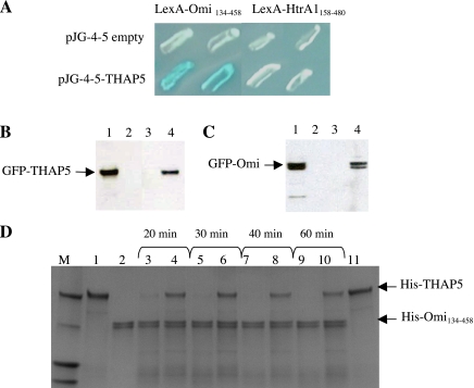
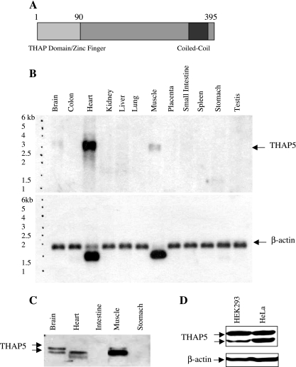
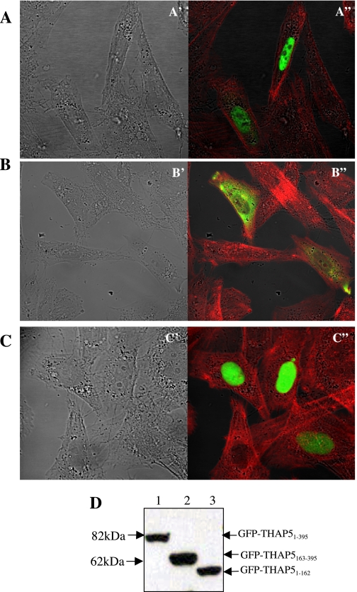
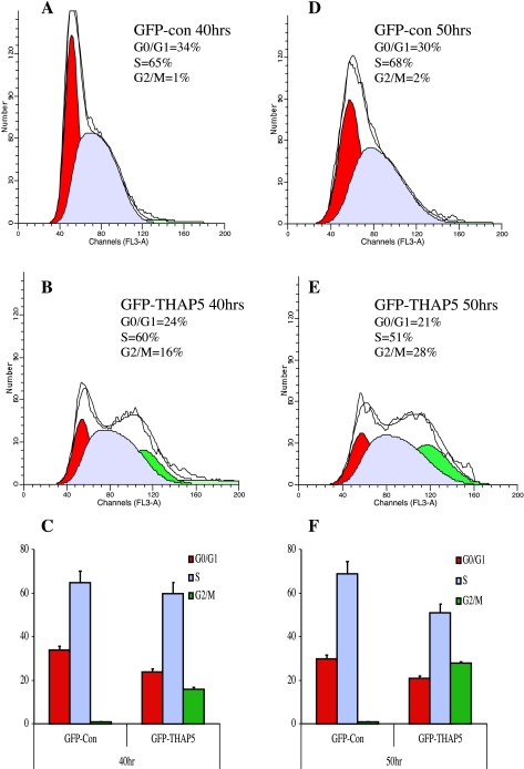
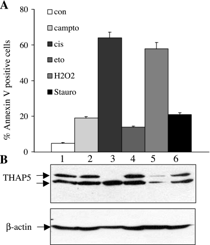
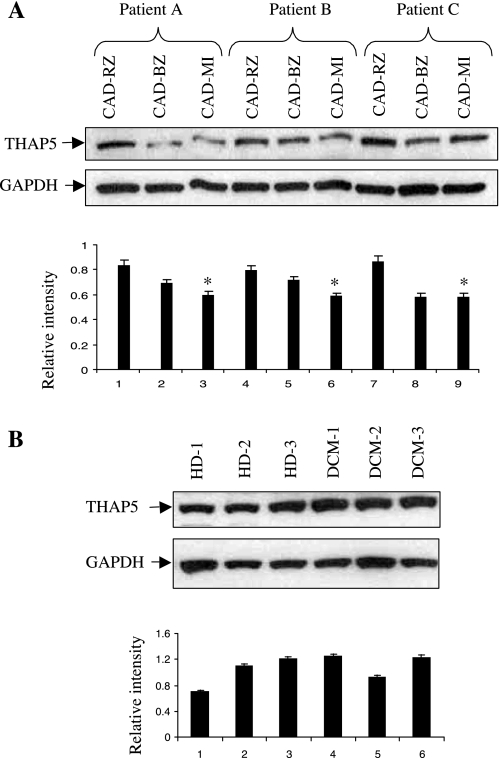
Omi/HtrA2 is a mitochondrial serine protease that has a dual function: while confined in the mitochondria, it promotes cell survival, but when released into the cytoplasm, it participates in caspase-dependent as well as caspase-independent cell death. To investigate the mechanism of Omi/HtrA2's function, we set out to isolate and characterize novel substrates for this protease. We have identified Thanatos-associated protein 5 (THAP5) as a specific interactor and substrate of Omi/HtrA2 in cells undergoing apoptosis. This protein is an uncharacterized member of the THAP family of proteins. THAP5 has a unique pattern of expression and is found predominantly in the human heart, although a very low expression is also seen in the human brain and muscle. THAP5 protein is localized in the nucleus and, when ectopically expressed, induces cell cycle arrest. During apoptosis, THAP5 protein is degraded, and this process can be blocked using a specific Omi/HtrA2 inhibitor, leading to reduced cell death. In patients with coronary artery disease, THAP5 protein levels substantially decrease in the myocardial infarction area, suggesting a potential role of this protein in human heart disease. This work identifies human THAP5 as a cardiac-specific nuclear protein that controls cell cycle progression. Furthermore, during apoptosis, THAP5 is cleaved and removed by the proapoptotic Omi/HtrA2 protease. Taken together, we provide evidence to support that THAP5 and its regulation by Omi/HtrA2 provide a new link between cell cycle control and apoptosis in cardiomyocytes.


Figures

Fig. 1.
Fig. 1.
Thanatos-associated protein (THAP) 5 is an interactor and substrate of Omi/HtrA2 protease. A: interaction of Omi/HtrA2 with THAP5 protein in yeast. Yeast colonies were transformed with plasmids encoding the indicated baits and full-length THAP5 cloned into the prey vector. The specificity of THAP5 and Omi/HtrA2 interaction was verified using a closely related homolog, the HtrA1 protein. Blue color results are from a positive protein-protein interaction. B: interaction of Omi/HtrA2 and THAP5 in mammalian cells during apoptosis. Human embryonic kidney (HEK)-293 cells were plated in duplicates in 60-mm dishes and then transfected with either enhanced green fluorescent protein (EGFP), EGFP-THAP5, or green fluorescent protein (GFP)-Omi. Twelve hours after transfection, one plate was treated with 50 μM cisplatin to induce apoptosis, and one plate was used as control. Cell lysates were prepared as described in materials and methods. A polyclonal Omi/HtrA2 antibody was used to immunoprecipitate the endogenous Omi/HtrA2 and any associated proteins. The immunoprecipitated complex was resolved on SDS-PAGE and transferred to a polyvinylidene difluoride (PVDF) membrane, and the presence of GFP-THAP5 fusion protein was detected using a specific anti-GFP antibody. Lane 1 shows crude lysates of GFP-THAP5 transfected cells. Lane 2 shows coimmunoprecipitation (co-IP) lysates obtained from cells transfected with GFP empty vector. Lane 3 shows co-IP lysates obtained from cells transfected with GFP-THAP5 control cells. Lane 4 shows co-IP lysates obtained from cells transfected with GFP-THAP5 and then treated with cisplatin. C: the reverse experiment described in B. HEK-293 cells were now transfected with GFP-Omi and processed exactly as in B, except THAP5 antiserum was used in the co-IP and anti-GFP on the Western blot to detect the GFP-Omi. Lane 1 shows total cell lysates of GFP-Omi transfected cells. Lane 2 shows co-IP lysates obtained from cells transfected with GFP empty vector. Lane 3 shows co-IP lysates obtained from cells transfected with GFP-Omi control cells. Lane 4 shows co-IP lysates obtained from cells transfected with GFP-Omi and then treated with cisplatin. In both B and C, THAP5 was coprecipitated with Omi/HtrA2, but only in cells in which apoptosis was induced. D: THAP5 is cleaved by Omi/HtrA2 protease in vitro. Purified His-THAP5 was incubated with His-Omi134-458 at 37°C for the indicated time periods. For some assays, Omi/HtrA2 was preincubated with ucf-101 inhibitor 10 min before the addition of His-THAP5. The reactions were resolved on SDS-PAGE, and the gel stained with Coomassie blue. Lane 1: His-THAP5 control (400 ng); lane 2: His-Omi134-458 control (400 ng); lanes 3, 5, 7, and 9: His-Omi+His-THAP5 at different time points; lane 4, 6, 8, and 10: His-Omi + His-THAP5 + ucf-101 (50 μM); lane 11: His-THAP5 + ucf-101 control.
Fig. 2.
Fig. 2.
Expression of THAP5: mRNA and protein in human tissues. A: schematic representation of the THAP5 protein encoded by 395 amino acids. The light gray box represents the 90aa THAP domain, which is characteristic of all THAP proteins. B: Northern blot analysis of THAP5 expression in human tissues. A commercially available Northern blot (Clontech) containing 2 μg/lane poly (A) mRNA from various adult human tissues was probed with 32P-THAP5 (539-1342) cDNA. A single transcript was detected of ∼3.2 kb. The blot clearly shows high expression of THAP5 in the human heart; some expression was also seen in skeletal muscle and brain. β-Actin probe was used to verify that equal amounts of mRNA were present in each lane. C: Western blot analysis of THAP5 protein in multiple human tissues. A commercial Western blot (INSTA-blot Calbiochem) containing 15 μg/lane of total protein from adult human tissues was incubated with rabbit-polyclonal anti-THAP5 antibody followed by a secondary antibody, horseradish peroxidase-conjugated goat anti-rabbit and chemiluminescence detection. D: Western blot analysis showing the relative expression of THAP5 in HEK-293 and HeLa cells. Thirty micrograms of whole cell lysates were resolved on SDS-PAGE, and the membrane was incubated with THAP5 polyclonal antibody. β-Actin antibody was used to verify that an equal amount of protein was loaded in each lane.
Fig. 3.
Fig. 3.
Subcellular localization of the THAP5 protein in mammalian cells. A: confocal image of HeLa cells transfected with GFP-THAP51-395 shows exclusive nuclear localization (green in A"). B: HeLa cells transfected with GFP-THAP5163-395 show cytoplasmic localization (green in B"). C: HeLa cells transfected with GFP-THAP51-162 show exclusive nuclear localization (green in C") of this protein. The cells were also stained with Texas red-phalloidin (Molecular Probes) that stain actin filaments (red). Nomarski/DIC images of the same cells are shown in A′, B′, and C′, respectively. D: the stability of EGFP-fusion proteins was monitored by Western blot analysis using an anti-GFP antibody. Equal amounts of whole cell lysates, obtained 24 h posttransfection, were subjected to SDS-PAGE followed by Western blot analysis using anti-GFP monoclonal antibody, as described in materials and methods. Lanes 1, 2, and 3 represent lysates obtained after transfection of HeLa cells with GFP-THAP51-395, GFP-THAP5163-395, and GFP-THAP51-162, respectively.
Fig. 4.
Fig. 4.
Overexpression of GFP-THAP5 causes accumulation of cells in G2/M phase. HEK-293 cells were transiently transfected with GFP vector alone or GFP-THAP5. Forty and fifty hours posttransfection, cells were stained with 7-amino-actinomycin D (7-AAD), and the DNA content was analyzed using ModFitLT software. The percentages of GFP positive cells at different phases of cell cycle 40 h (C) and 50 h (F) after transfection are shown. Histogram results are shown from representative experiments in cells transfected with GFP vector at 40 h (A) and 50 h (D). Cells transfected with GFP-THAP5 for 40 h (B) and 50 h (E) show significant increase in the percentage of cells in G2/M phase. Data are means ± SD of four independent experiments.
Fig. 5.
Fig. 5.
THAP5 protein level is regulated during apoptosis. Total cell lysates were prepared from HeLa cells after induction of apoptosis using different chemicals (camptothecin, cisplatin, etoposide, H2O2, and staurosporine). A: cell death was monitored in the treated cell populations by annexin V and 7-AAD staining and analyzed by flow cytometry. B: cell extracts were prepared followed by Western blot analysis of the same samples. Lane 1 shows lysates from control, untreated cells; lanes 2, 3, and 4 show cell extracts from HeLa treated with the chemical indicated above. β-Actin antibody was used to verify that equal amounts of protein were present in each lane. Data are means ± SD of four independent experiments.
Fig. 6.
Fig. 6.
A specific inhibitor of Omi/HtrA2 blocks THAP5 cleavage and protects HeLa cells from apoptosis. A: HeLa cells were treated with 20 or 30 μM of ucf-101, and apoptosis was induced with 5 μM cisplatin for 12 h. Apoptosis was monitored using annexin V and 7-AAD staining and analyzed by flow cytometry. B: extracts were prepared from the same cell population and analyzed by SDS-PAGE and Western blot analysis using THAP5 antibody. Cisplatin treatment caused a dramatic reduction in THAP5 protein level (lane 2). This corresponds with increased apoptosis in the cell population. When HeLa cells were treated with ucf-101 followed by cisplatin, the inhibitor substantially protected THAP5 proteins, and the percentage of apoptotic cells was significantly reduced (lanes 3 and 4). β-Actin antibody was used to verify that equal amounts of protein were present in each lane. Data are means ± SD of three independent experiments.
Fig. 7.
Fig. 7.
THAP5 protein levels in heart tissues from patients with coronary artery disease (CAD). Heart tissues were obtained from patients with end-stage heart failure due to CAD or dilated cardiomyopathy (DCM) and were undergoing cardiac transplantation. A: heart tissue extracts from three patients with CAD were used in a Western blot to monitor the expression of THAP5 protein. Top: extracts were prepared from three areas on each heart corresponding to the remote zone (CAD-RZ), border zone (CAD-BZ), and the infarction (CAD-MI) area. Bottom: THAP5-to-GAPDH ratios were calculated after densitometry. *P < 0.05 vs. CAD-RZ. B, top: human heart tissue lysates from three healthy donors (HD-1, -2, and -3) and three DCM patients (DCM-1, -2, and -3) were probed for THAP5 expression. Data are means ± SD (n = 6 patients/group). Bottom: THAP5-to-GAPDH ratios were calculated after densitometry.

References

    1. Adachi S, Ito H, Tamamori-Adachi M, Ono Y, Nozato T, Abe S, Ikeda M, Marumo F, Hiroe M. Cyclin A/cdk2 activation is involved in hypoxia-induced apoptosis in cardiomyocytes. Circ Res 88: 408–414, 2001. - PubMed
    1. Bhuiyan MS, Fukunaga K. Activation of HtrA2, a mitochondrial serine protease mediates apoptosis: current knowledge on HtrA2 mediated myocardial ischemia/reperfusion injury. Cardiovasc Ther 26: 224–232, 2008. - PubMed
    1. Bhuiyan MS, Fukunaga K. Inhibition of HtrA2/Omi ameliorates heart dysfunction following ischemia/reperfusion injury in rat heart in vivo. Eur J Pharmacol 557: 168–177, 2007. - PubMed
    1. Boxem M, van den Heuvel S. C. elegans class B synthetic multivulva genes act in G(1) regulation. Curr Biol 12: 906–911, 2002. - PubMed
    1. Boxem M, van den Heuvel S. lin-35 Rb and cki-1 Cip/Kip cooperate in developmental regulation of G1 progression in C. elegans. Development 128: 4349–4359, 2001. - PubMed

Publication types

MeSH terms