. 2009 Jul;16(7):763-8.
doi: 10.1038/nsmb.1611. Epub 2009 Jun 7.

ATXR5 and ATXR6 are H3K27 monomethyltransferases required for chromatin structure and gene silencing

Affiliations

ATXR5 and ATXR6 are H3K27 monomethyltransferases required for chromatin structure and gene silencing

Yannick Jacob et al. Nat Struct Mol Biol. 2009 Jul.

Abstract

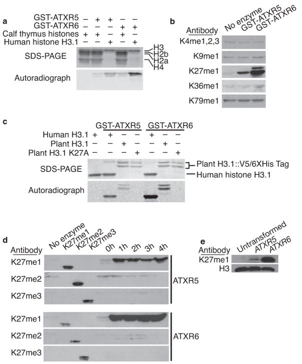
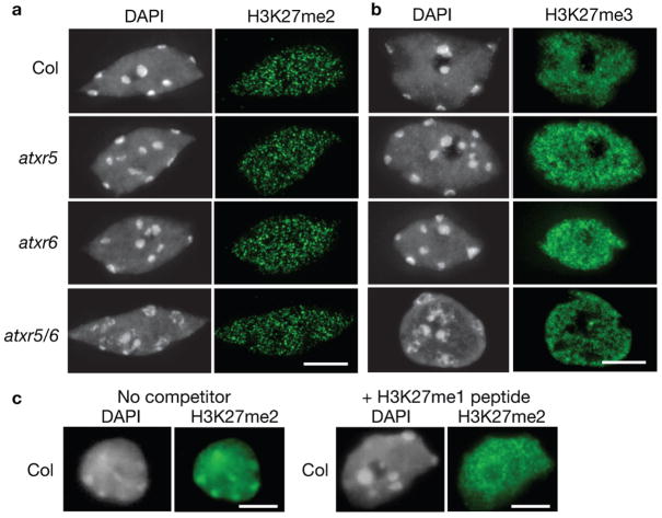
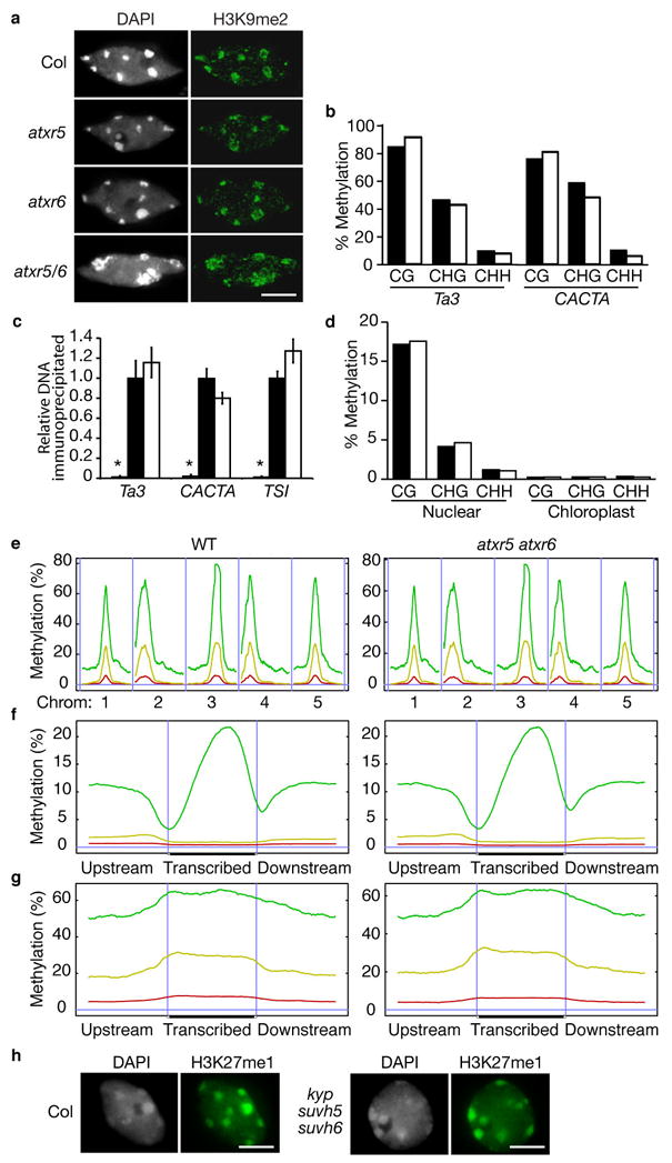
Constitutive heterochromatin in Arabidopsis thaliana is marked by repressive chromatin modifications, including DNA methylation, histone H3 dimethylation at Lys9 (H3K9me2) and monomethylation at Lys27 (H3K27me1). The enzymes catalyzing DNA methylation and H3K9me2 have been identified; alterations in these proteins lead to reactivation of silenced heterochromatic elements. The enzymes responsible for heterochromatic H3K27me1, in contrast, remain unknown. Here we show that the divergent SET-domain proteins ARABIDOPSIS TRITHORAX-RELATED PROTEIN 5 (ATXR5) and ATXR6 have H3K27 monomethyltransferase activity, and atxr5 atxr6 double mutants have reduced H3K27me1 in vivo and show partial heterochromatin decondensation. Mutations in atxr5 and atxr6 also lead to transcriptional activation of repressed heterochromatic elements. Notably, H3K9me2 and DNA methylation are unaffected in double mutants. These results indicate that ATXR5 and ATXR6 form a new class of H3K27 methyltransferases and that H3K27me1 represents a previously uncharacterized pathway required for transcriptional repression in Arabidopsis.


Conflict of interest statement

COMPETING INTEREST STATEMENT

The authors declare that they have no competing financial interests.

Figures

Figure 1
Figure 1
ATXR5 and ATXR6 monomethylate H3K27. (a) In vitro histone methyl transferase assay using radiolabeled SAM; only H3 is methylated by ATXR5 and ATXR6. (b) The products of non-radioactive HMT assays were analyzed by Western blot using antibodies against different methylated lysines of H3. (c) ATXR5 and ATXR6 cannot methylate a mutant plant H3 protein where lysine 27 has been replaced with alanine. (d) ATXR5 and ATXR6 HMT time-course assay products were analyzed by Western blot using antibodies specific for mono-, di-, and trimethylated forms of H3K27. H3K27 methylated peptides were included to confirm the specificity of the antibodies and a no-enzyme negative control was included. (e) Western blot analysis of histones extracted from S. cerevisiae expressing ATXR5 and ATXR6.
Figure 2
Figure 2
ATXR5 and ATXR6 play redundant roles in leaf development. (a) Schematic drawings indicate the genomic structure of ATXR5 and ATXR6; thin horizontal lines represent the chromosome, thick lines represent exons. Horizontal lines below the drawings represent the regions amplified by each primer pair. (b, c) Semi-quantitative RT-PCR analysis of ATXR5 and ATXR6 expression in (b) fully expanded leaves and (c) various tissues. UBQ was used as a constitutively expressed control. (d) Single and double mutants of atxr5 and atxr6 grown under short-day conditions. (e) Rate of leaf initiation of plants were grown under short days.
Figure 3
Figure 3
atxr5 atxr6 mutations lead to disruption of constitutive heterochromatin, reduced H3K27 monomethylation, and reactivation of silenced elements. (a) Leaf interphase nuclei were stained with DAPI and analyzed for immunofluorescence with an anti-H3K27me1 antibody. Approximately 65% of atxr5 atxr6 nuclei show severe (a, upper panel) or moderate (a, lower panel) chromocenter decondensation and reduced H3K27me1 staining. Scale bar = 5 μm. (b) FISH analysis of leaf interphase nuclei using a 180-bp centromeric repeat probe. The DNA was counterstained with DAPI. Scale bar = 5 μm. (c) Semi-quantitative RT-PCR analysis of heterochromatic elements in Col, atxr5, atxr6, and atxr5 atxr6. UBQ was used as a constitutively expressed control. (d) ChIP analysis of repetitive elements using H3K27me1 antibodies. Black and white bars indicate relative levels of immunoprecipitated DNA normalized to ACTIN, as determined by real-time PCR, from wild type and atxr5 atxr6 leaves, respectively. Grey bars represent no antibody controls. The data are presented as mean ± SEM for three individual experiments.
Figure 4
Figure 4
Di- and trimethylation of H3K27 are not altered in atxr5 atxr6 mutants (a, b) Leaf interphase nuclei were stained with DAPI and analyzed for immunoflorescence with (a) anti-H3K27me2 and (b) anti-H3K27me3 antibodies. c, Leaf interphase nuclei were stained with DAPI and analyzed for immunofluorescence with anti-H3K27me2 in the presence or absence of an H3K27me1 peptide. Scale bars = 5 μm.
Figure 5
Figure 5
Mutations in atxr5 and atxr6 do not affect H3K9 dimethylation or DNA methylation. (a) Leaf interphase nuclei were stained with DAPI and analyzed for immunoflorescence with anti-H3K9me2 antibodies. Scale bar = 5 μm. (b, d) DNA methylation analysis by (b) locus-specific (Ta3 and CACTA) and (d) genome-wide BS-Seq. Black and white bars represent wild type and atxr5 atxr6, respectively. (c) ChIP analysis of repetitive elements using H3K9me2 antibodies. Black and white bars indicate relative levels of immunoprecipitated DNA normalized to ACTIN, as determined by real-time PCR, from wild type and atxr5 atxr6, respectively. Grey bars represent no antibody controls. The data are presented as mean ± SEM for three individual experiments. (e) Distribution of methylation along the five Arabidopsis chromosomes. (f) Average methylation levels within protein coding genes. (g) Average methylation levels within pseudogenes and transposons. (e–g) A horizontal blue line indicates zero percent methylation. Panels on the left correspond to wild type and panels on the right correspond to atxr5 atxr6 double mutant. CG methylation is indicated by green lines, CHG methylation is indicated by yellow lines, and CHH methylation is indicated by red lines. (e) Vertical blue lines are used to separate different chromosomes. (f, g) Vertical blue lines mark the boundaries between upstream regions and gene bodies and between gene bodies and downstream regions. (h) Leaf interphase nuclei of wild-type plants and kyp suvh5 suvh6 triple mutants were stained with DAPI and analyzed for immunofluorescence with anti-H3K27me1. Scale bar = 5 μm.

References

    1. Li B, Carey M, Workman JL. The role of chromatin during transcription. Cell. 2007;128:707–19. - PubMed
    1. Maluszynska J, Heslop-Harrison JS. Localization of tandemly repeated DNA sequences in Arabidopsis thaliana. Plant J. 1991;1:159–166.
    1. Cokus SJ, et al. Shotgun bisulphite sequencing of the Arabidopsis genome reveals DNA methylation patterning. Nature. 2008;452:215–9. - PMC - PubMed
    1. Johnson L, Cao X, Jacobsen S. Interplay between two epigenetic marks. DNA methylation and histone H3 lysine 9 methylation. Curr Biol. 2002;12:1360–7. - PubMed
    1. Probst AV, Fransz PF, Paszkowski J, Mittelsten Scheid O. Two means of transcriptional reactivation within heterochromatin. Plant J. 2003;33:743–9. - PubMed

Publication types

Associated data