. 2009 Jul;12(7):857-63.
doi: 10.1038/nn.2334. Epub 2009 Jun 7.

NADPH oxidase is the primary source of superoxide induced by NMDA receptor activation

Affiliations

NADPH oxidase is the primary source of superoxide induced by NMDA receptor activation

Angela M Brennan et al. Nat Neurosci. 2009 Jul.

Abstract

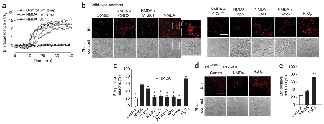
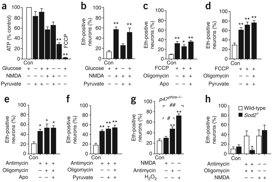
Neuronal NMDA receptor (NMDAR) activation leads to the formation of superoxide, which normally acts in cell signaling. With extensive NMDAR activation, the resulting superoxide production leads to neuronal death. It is widely held that NMDA-induced superoxide production originates from the mitochondria, but definitive evidence for this is lacking. We evaluated the role of the cytoplasmic enzyme NADPH oxidase in NMDA-induced superoxide production. Neurons in culture and in mouse hippocampus responded to NMDA with a rapid increase in superoxide production, followed by neuronal death. These events were blocked by the NADPH oxidase inhibitor apocynin and in neurons lacking the p47(phox) subunit, which is required for NADPH oxidase assembly. Superoxide production was also blocked by inhibiting the hexose monophosphate shunt, which regenerates the NADPH substrate, and by inhibiting protein kinase C zeta, which activates the NADPH oxidase complex. These findings identify NADPH oxidase as the primary source of NMDA-induced superoxide production.


Figures

Figure 1
Figure 1
NADPH oxidase is the major source of NMDA-induced superoxide formation in neurons. (a) Traces show ethidium (eth) fluorescence over time, as measured in individual neurons preloaded with dHEth. Following baseline fluorescence recording, neurons were treated with 100 µM NMDA (arrow) at either 21–25 °C or 35 °C. Traces are representative of >40 neurons under each condition from four independent studies. (b) Ethidium fluorescence and corresponding phase contrast images of cortical neuron cultures photographed 30 min after incubation with NMDA (100 µM) alone or with MK801 (10 µM), CNQX (10 µM), apocynin (apo, 500 µM), 6AN (500 µM) or Trolox (100 µM). For calcium-free experiments, neurons were incubated with a zero-calcium buffer and 1 mM EGTA. Control wells received medium exchanges only. H2O2 (100 µM) served as a positive control. Inset, higher magnification of ethidium-positive neurons from the region indicated by the white box. Scale bar represents 10 µm. (c) Quantification of ethidium-positive neurons under the conditions shown in b. (d) Ethidium fluorescence and corresponding phase-contrast images of p47phox−/− neurons incubated with NMDA (100 µM) or H2O2 (100 µM). Scale bar represents 10 µm. (e) Quantification of ethidium-positive neurons under the conditions shown in d. * P < 0.05 and ** P < 0.01 versus NMDA alone (n ≥ 3). Error bars indicate s.e.m.
Figure 2
Figure 2
NMDA-induced oxidative damage was prevented by NADPH oxidase inhibition. (a–d) Immunostaining for 4HNE in wild-type (a) and p47phox−/− (c) neuron cultures (conditions as described in Fig. 1). 4HNE immunostaining is quantified in b and d for a and c, respectively. * P < 0.05 and ** P < 0.01 versus control (n = 3). Scale bars represent 10 µm. Error bars indicate s.e.m.
Figure 3
Figure 3
Mitochondria do not substantially contribute to NMDA-induced superoxide production. (a) We found comparable ATP levels in neurons incubated with 5 mM glucose or 1 mM pyruvate, confirming that mitochondrial respiration is supported by pyruvate. ATP levels were reduced by the absence of both substrates or by the presence of FCCP (3 µM). (b) Glucose was required for NMDA-induced superoxide production and this was not suppressed by pyruvate (500 µM). (c–g) The ethidium fluorescence method detected mitochondrial superoxide produced by neurons incubated with FCCP (3 µM, c,d) or with the complex III inhibitor antimycin (3 µM, e,f), with or without 3 µM oligomycin. This mitochondrial ethidium signal was not attenuated by 500 µM apocynin or pyruvate (c–f) or in p47phox−/− neurons (g). (h) Neurons prepared from Sod2+ mice had an attenuated ethidium response to antimycin and oligomycin relative to wild-type littermates, but had no reduction in ethidium increase following NMDA application. * P < 0.05 and ** P < 0.01 versus control, # P < 0.05 and ## P < 0.01 versus NMDA (n ≥ 3). Error bars indicate s.e.m.
Figure 4
Figure 4
NMDA activation of NADPH oxidase is mediated by PKC. (a) Ethidium fluorescence and corresponding phase-contrast images of cortical neuron cultures photographed 30 min after incubation with NMDA (100 µM) alone or with the peptide inhibitors PKCδ, PKCζ, or TAT carrier peptide (all 1 µM). Scale bar represents 10 µm. (b) Quantification of Eth fluorescence. (c) Immunostaining for p47phox and MAP2 in neurons indicated that NMDA-induced translocation of the p47phox subunit to the cell membrane was attenuated by the PKCζ peptide inhibitor. Scale bar represents 2 µm. (d) Quantification of p47phox membrane translocation. * P < 0.05 versus NMDA and ** P < 0.05 versus control (n ≥ 4). Error bars indicate s.e.m.
Figure 5
Figure 5
Inhibition of NADPH oxidase prevents NMDA-induced cell death. (a–f) Dead neurons were identified with trypan blue staining 24 h after NMDA incubations. 0 glu + pyr indicates glucose-free medium with 1 mM pyruvate. All other conditions are as described in Figure 1 and Figure 3. Neuronal death is quantified in b, d and f for a, c and e, respectively. Scale bar = 40 µm. * P < 0.05 and ** P < 0.01 versus NMDA, *** P < 0.05 versus control, and † P < 0.05 versus wild-type NMDA (n = 4). Error bars indicate s.e.m.
Figure 6
Figure 6
NMDA induced superoxide in mouse hippocampus. (a) Representative images of ethidium fluorescence in neurons from the CA1 hippocampus of wild-type or p47phox−/− mice. Tissue was harvested 30 min after NMDA injections. (b) Comparison of ethidium intensity between groups. (c) Representative images of NMDA-induced ethidium fluorescence in neurons from the CA1 hippocampus of wild-type mice pre-treated with peptide inhibitors of PKCδ and PKCζ or the TAT carrier peptide. (d) Quantification of ethidium intensity between groups. (e) Representative images of degenerating neurons, stained with Fluoro-Jade B in the CA1 hippocampus of wild-type or p47phox−/− mice 3 d after NMDA injections. (f) Quantification of Fluoro-Jade B–positive neurons. Scale bars represent 50 µm. * P < 0.05 (n = 4). Error bars indicate s.e.m.
Figure 7
Figure 7
NMDA-induced superoxide production in cell bodies and dendrites. Confocal images of mouse CA1 hippocampus after immunostaining for the neuronal marker MAP2 and the lipid oxidation product 4HNE. Brains were harvested 30 min after stereotactic injections with NMDA. 4HNE formation was evident in the neuronal cell bodies of the stratum pyramidale (SP) and in the dendrites extending from the stratum radiatum (SR). 4HNE formation was markedly reduced in p47phox−/− mice. Scale bar represents 40 µm. Images are representative of three mice from each group.

Comment in

References

    1. Lafon-Cazal M, Pietri S, Culcasi M, Bockaert J. NMDA-dependent superoxide roduction and neurotoxicity. Nature. 1993;364:535–537. - PubMed
    1. Mandir AS, et al. NMDA, but not non-NMDA, excitotoxicity is mediated by poly(ADP-ribose) polymerase. J. Neurosci. 2000;20:8005–8011. - PMC - PubMed
    1. MacDermott AB, Mayer ML, Westbrook GL, Smith SJ, Barker JL. NMDA receptor activation increases cytoplasmic calcium concentration in cultured spinal cord neurones. Nature. 1986;321:519–522. - PubMed
    1. Klann E. Cell-permeable scavengers of superoxide prevent long-term potentiation in hippocampal area CA1. J. Neurophysiol. 1998;80:452–457. - PubMed
    1. MacDonald JF, Jackson MF, Beazely MA. Hippocampal long-term synaptic lasticity and signal amplification of NMDA receptors. Crit. Rev. Neurobiol. 2006;18:71–84. - PubMed

Publication types

MeSH terms

LinkOut - more resources