

doi: 10.1364/opex.13.010217.
High speed spectral domain polarization sensitive optical coherence tomography of the human retina
- PMID: 19503236
- PMCID: PMC2978948
- DOI: 10.1364/opex.13.010217
Item in Clipboard
High speed spectral domain polarization sensitive optical coherence tomography of the human retina
Opt Express.
.
Abstract
We developed a high-speed polarization sensitive optical coherence tomography (PS-OCT) system for retinal imaging based on spectral domain OCT. The system uses two spectrometers, one for each polarization channel, that operate in parallel at 20000 A-lines/s each. It provides reflectivity, retardation, and cumulative optic axis orientation simultaneously. We present our instrument and discuss the requirements for the alignment of the two spectrometers specific for our setup. We show 2D spectral domain PS-OCT images and - to the best of our knowledge - the first 3D spectral domain PS-OCT data sets in form of fly-through movies and volume rendered data sets recorded in human retina in vivo.
Figures
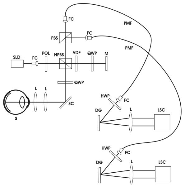
Schematic drawing of spectral domain PS-OCT instrument. SLD, super luminescent diode; FC, fiber coupler; POL, polarizer; NPBS, non-polarizing beam splitter; VDF, variable density filter; QWP, quarter wave plate; M, mirror; SC, galvo scanner; L, lens; S, sample; PMF, polarization maintaining fiber; HWP, half wave plate; DG, diffraction grating; LSC, line scan camera.

Results of instrument calibration. A sample consisting of a wave plate and mirror was measured. Axis orientation and retardation are plotted as a function of path length difference. Within the desired measurement range of 2 mm, the axis drift stays within ± 3°.
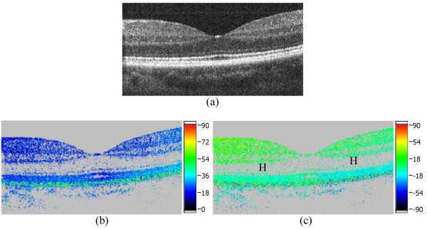
B-scan images of human fovea in vivo. (a) Intensity (log scale); (b) retardation; (c) fast axis orientation. Image size: 3 mm (horizontal) × 0.75 mm (vertical). Values on color bars: degrees (to avoid erroneous birefringence data, areas below a certain intensity threshold are displayed in grey).

B-scan images of human optic nerve head in vivo. (a) Intensity (log scale); (b) retardation; (c) fast axis orientation. Image size: 3 mm (horizontal) × 1.75 mm (vertical). Values on color bars: degrees. Arrow: temporal rim of scleral canal.

B-scans superior to human optic nerve head in vivo. (a) Intensity (log scale); (b) retardation; (c) fast axis orientation. Image size: 3 mm (horizontal) × 1 mm (vertical). Values on color bars: degrees.

(2.5 MB) Frame no. 31 of fly-through movie of 3D dataset of human nerve head in vivo. Top: intensity; middle: retardation; bottom: axis orientation (color scales similar to Figs. 2-4). Image size: ~ 3mm (x) × 3mm (y) × 1.75mm (z).

(1.2 MB) Frame no. 4 of animation of a 3 dimensional volume rendered data set from a human nerve head in vivo. The opacity corresponds to the backscattered intensity, the retardation corresponds to the color coding (color coding similar to Figs. 2-4).

(2.2 MB) Frame no. 3 of animation of a 3 dimensional volume rendered data set from a human nerve head in vivo. The opacity corresponds to the backscattered intensity, the fast axis orientation corresponds to the color coding (color coding similar to Figs. 2-4).

PS-OCT axis orientation image of fovea centralis in vivo. The image illustrates the effect of translationally misaligned spectrometer cameras. Image size: ~ 3 mm (horizontal) × 0.75 mm (vertical).
References
-
- Bouma BE, Tearney GJ. Handbook of optical coherence tomography. Marcel Dekker; New York: 2002.
-
- Fercher AF, Hitzenberger CK. Optical Coherence Tomography. Prog. Opt. 2002;44:215–302.
-
- Hee MR, Huang D, Swanson EA, Fujimoto JG. Polarization sensitive low coherence reflectometer for birefringence characterization and ranging. J. Opt. Soc. Am. B. 1992;9:903–908.
-
- de Boer JF, Milner TE, Van Gemert MJC, Nelson JS. Two-dimensional birefringence imaging in biological tissue by polarization-sensitive optical coherence tomography. Opt. Lett. 1997;22:934–936. - PubMed
Grants and funding
LinkOut - more resources
Full Text Sources
Other Literature Sources
