

A case of subretinal neovascularization treated with intravitreal ranibizumab in a patient with idiopathic juxtafoveal retinal telangiectasis
- PMID: 19503767
- PMCID: PMC2685226
A case of subretinal neovascularization treated with intravitreal ranibizumab in a patient with idiopathic juxtafoveal retinal telangiectasis
Abstract
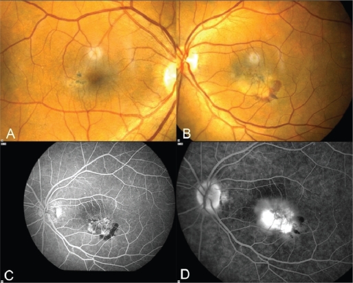
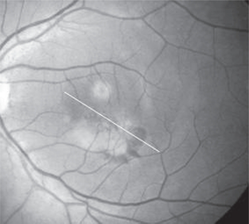
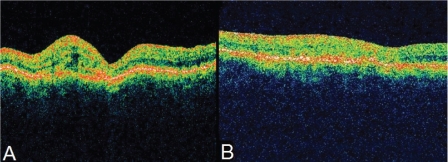
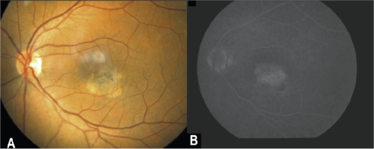
A 65-year-old lady presented with decreased vision in her left eye (LE). Best corrected visual acuity (BCVA) was 1/20. Complete examination showed idiopathic juxtafoveal retinal telangiectasis associated with subretinal neovascularization and she was treated with intravitreal ranibizumab every month for three months in the LE. After four months, her BCVA increased to 3/10. Fluorescein angiography (FA) showed minimal leakage and optical coherence tomography (OCT) confirmed absence of intra- or subretinal fluid in the macula. Examinations were repeated monthly for another 12 months and showed no recurrence. Intravitreal ranibizumab showed promising results for subretinal neovascularization due to idiopathic juxtafoveal retinal telangiectasis. A prospective study with large series of patients and controls may be necessary in order to determine the effectiveness of this treatment.
Keywords: idiopathic juxtafoveal retinal telangiectasis; ranibizumab; subretinal neovascularization.
Figures
References
-
- Gass JD, Blodi BA. Idiopathic juxtafoveal retinal telangiectasis. Update of classification and follow-up study. Ophthalmology. 1993;100:1536–1546. - PubMed
-
- Engelbrecht NE, Aaberg TM, Jr, Sung J, Lewis ML. Neovascular membranes associated with idiopathic juxtafoveolar telangiectasis. Arch Ophthalmol. 2002;120:320–324. - PubMed
-
- Yannuzzi LA, Bardal AM, Freund KB, Chen KJ, Eandi CM, Blodi B. idiopathic macular telangiectasia. Arch Ophthalmol. 2006;124:450–460. - PubMed
-
- Snyers B, Verougstraete C, Postelmans L, Leys A, Hykin P. Photodynamic therapy of subfoveal neovascular membrane in type 2A idiopathic juxtafoveolar retinal telangiectasis. Am J Ophthalmol. 2004;137:812–819. - PubMed
-
- Charbel Issa P, Finger RP, Holz FG, Scholl HP. Eighteen-month follow-up of intravitreal bevacizumab in type 2idiopathic macular telangiectasia. Br J Ophthalmol. 2008;92:941–945. - PubMed
Publication types
MeSH terms
Substances
LinkOut - more resources
Full Text Sources
