. 2009 Jun 8;4(6):e5824.
doi: 10.1371/journal.pone.0005824.

Fgf and Sdf-1 pathways interact during zebrafish fin regeneration

Affiliations

Fgf and Sdf-1 pathways interact during zebrafish fin regeneration

Mohamed Bouzaffour et al. PLoS One. .

Abstract

The chemokine stromal cell-derived factor-1 (SDF1) was originally identified as a pre-B cell stimulatory factor but has been recently implicated in several other key steps in differentiation and morphogenesis. In addition, SDF1 as well as FGF signalling pathways have recently been shown to be involved in the control of epimorphic regeneration. In this report, we address the question of a possible interaction between the two signalling pathways during adult fin regeneration in zebrafish. Using a combination of pharmaceutical and genetic tools, we show that during epimorphic regeneration, expression of sdf1, as well as of its cognate receptors, cxcr4a, cxcr4b and cxcr7 are controlled by FGF signalling. We further show that, Sdf1a negatively regulates the expression of fgf20a. Together, these results lead us to propose that: 1) the function of Fgf in blastema formation is, at least in part, relayed by the chemokine Sdf1a, and that 2) Sdf1 exerts negative feedback on the Fgf pathway, which contributes to a transient expression of Fgf20a downstream genes at the beginning of regeneration. However this feedback control can be bypassed since the Sdf1 null mutants regenerate their fin, though slower. Very few mutants for the regeneration process were isolated so far, illustrating the difficulty in identifying genes that are indispensable for regeneration. This observation supports the idea that the regeneration process involves a delicate balance between multiple pathways.


Conflict of interest statement

Competing Interests: The authors have declared that no competing interests exist.

Figures

Figure 1
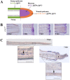
Figure 1. Expression of sdf1 and its receptors during fin regeneration.
A: schematic representation of FGF and SDF pathways in caudal fin regeneration. In green: stump epidermis expressing cxcr4a and fgfr1. In red: wound epidermis expressing cxcr4b, cxcr7, fgf20a and fgfr1. In blue: blastema expressing sdf1a, fgf20a and fgfr1. Longitudinal cross section through the dermal ray of a regenerating fin at 2 dpa. The dotted line indicates the amputation plane. B: cxcr7 kinetics of expression during caudal fin regeneration. cxcr7 mRNA expression pattern was analyzed by in situ hybridization on control fin (0 dpa) and in amputated fins allowed to regenerate for 2, 3 and 4 dpa. Scale bar, 100 µm. C: In situ hybridization for cxcr7 on cryosections. cxcr7 mRNA expression pattern was analyzed by in situ hybridization on cryosections of uncut fin (0 dpa) and in amputated fins allowed to regenerate for 2 dpa. Two days post amputation cxcr7 mRNA is detected in the wound epidermis as well as in a few dispersed cells in the stump epidermis (enlarged view). Before amputation, only few mesenchymal cells show a staining for cxcr7. Scale bars, 50 µm.
Figure 2
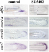
Figure 2. FgfR inhibition modifies sdf1a, cxcr4a, cxcr4b and cxcr7 expression in ongoing fin regenerates.
Sections of 2 dpa caudal fins from fish treated with DMSO (control) or FGFR inhibitor (SU5402) after in situ hybridization for sdf1a, cxcr4a, cxcr4b and cxcr7. Scale bar, 100 µm.
Figure 3
Figure 3. SDF1 inhibits fgf20a and activates Wnt10a expression during fin regeneration.
A: Over-expression of SDF1 at the time of amputation turns off fgf20a expression. The protein SDF1, or BSA as a control, were injected into the fin at the time of amputation. Fins were allowed to regenerate for 48 hours before fgf20a expression was checked by ISH. Dotted lines demarcate amputation plane. Scale bar, 100 µm. B: fgf20a expression is enhanced in medusa mutant. fgf20a as well as wnt10a expression were analyzed by quantitative RT-PCR in sdf1a/medusa mutant and sibling fins at 48 hpa. fgf20a expression is increased 2.2±0.03 fold in sdf1 mutant compared to siblings, while wnt10a expression is reduced 1.5±0.04 fold. Average values (±s.e.m.) from experiments performed in triplicate are presented. C, D: Fins regenerate slower in sdf1/medusa mutant compared to siblings (C) whereas cxcr4b mutant fish regenerates caudal fin with no abnormality (D). Length of the third dorsal and ventral regenerating fin ray of the regenerates were measured in ody−/− mutant fish and siblings (ody+/− and ody+/+) (D) and in medusa/sdf1−/− mutant fish and siblings (sdf1+/+ and sdf1+/−) (C) at 4 and 7 dpa. Precise measures were made in pixels (1 pixel corresponding to 3 µm). The average length of the regenerate allows to calculate the regeneration speed. No significant difference was observed between ody−/− mutant fish and siblings. Two independent experiments were performed. Experiment 1: n = 5 ody−/−, n = 7 siblings. Experiment 2 n = 5 ody−/−, n = 7 siblings (data not shown). Errors bars represent the s.e.m. of the average regenerate length. Fins regenerate slower in sdf1/medusa mutant n = 18 siblings (sdf1+/+ and sdf1+/−), n = 18 medusa/sdf1−/−. Errors bars represent the s.e.m. of the average regenerate length (* p<0.05). E–F: cxcr7 overexpression inhibits blastema formation. Plasmid DNA expressing cxcr7 (pCS2-cxcr7) was injected into the dorsal half fin whereas an empty plasmid (pCS2) was injected into the ventral part of the fin at the time of amputation prior to electroporation. Fins were allowed to regenerate for 48 hours before scoring blastema formation (n = 8). The percent area of dorsal versus ventral regrowth is presented in E and a representative fin in F. Scale bar, 500 µm. Dotted lines demarcate amputation plane. G: cxcr7 overexpression inhibits fgf20 expression. Plasmid DNA expressing cxcr7 (pCS2-cxcr7) or an empty plasmid (pCS2) as control were electroporated into total fin at the time of amputation. Fins were allowed to regenerate for 48 hours before checking fgf20 expression by quantitative PCR. Two independent experiments are presented in G. Experiment 1: n = 5; experiment 2: n = 5.
Figure 4
Figure 4. A model for signalling events regulating initiation of regeneration and fin outgrowth.
Amputation of the caudal fin triggers wnt10a expression which then activates fgf20 in a β-catenin-dependent mechanism . Fgf signalling (most likely through FgfR1) activates the expression of the chemokine sdf1 and its receptors cxcr4a and b, and then inhibits cxcr7 expression. In return Sdf1a exerts a negative feedback on fgf20a expression in parallel to Wnt5b. We propose that as soon as the level of Fgf20a protein decreases, this allows cxcr7 expression, Cxcr7 subsequently down regulating fgf20a in parallel to Wnt 5b. In this context the SDF1 pathway amplifies the negative regulation of fgf20a expression. In summary, Sdf1 thus plays two roles in fin regeneration. First it mediates Fgf activity to promote proliferation of epithelial cells , second it subsequently switches off Fgf activity by down regulating fgf20 expression.

References

    1. Nakatani Y, Kawakami A, Kudo A. Cellular and molecular processes of regeneration, with special emphasis on fish fins. Dev Growth Differ. 2007;49:145–154. - PubMed
    1. Poss KD, Keating MT, Nechiporuk A. Tales of regeneration in zebrafish. Dev Dyn. 2003;226:202–210. - PubMed
    1. Akimenko MA, Mari-Beffa M, Becerra J, Geraudie J. Old questions, new tools, and some answers to the mystery of fin regeneration. Dev Dyn. 2003;226:190–201. - PubMed
    1. Slack JM, Tannahill D. Mechanism of anteroposterior axis specification in vertebrates. Lessons from the amphibians. Development. 1992;114:285–302. - PubMed
    1. Slack J. Role of fibroblast growth factors as inducing agents in early embryonic development. Mol Reprod Dev. 1994;39:118–124. discussion 124–115. - PubMed

Publication types