. 2009 Jun 8;4(6):e5837.
doi: 10.1371/journal.pone.0005837.

Endotoxin induced chorioamnionitis prevents intestinal development during gestation in fetal sheep

Affiliations

Endotoxin induced chorioamnionitis prevents intestinal development during gestation in fetal sheep

Tim G A M Wolfs et al. PLoS One. .

Abstract

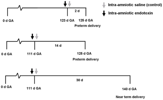
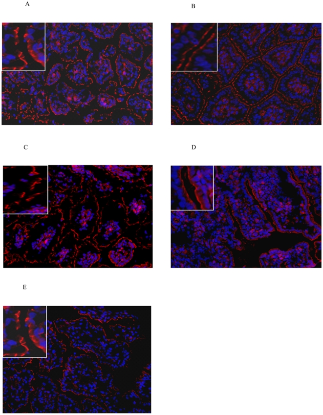
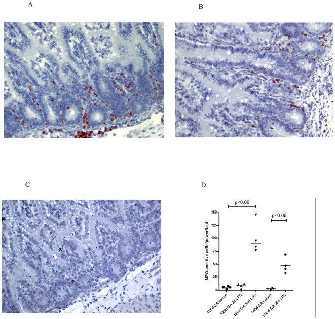
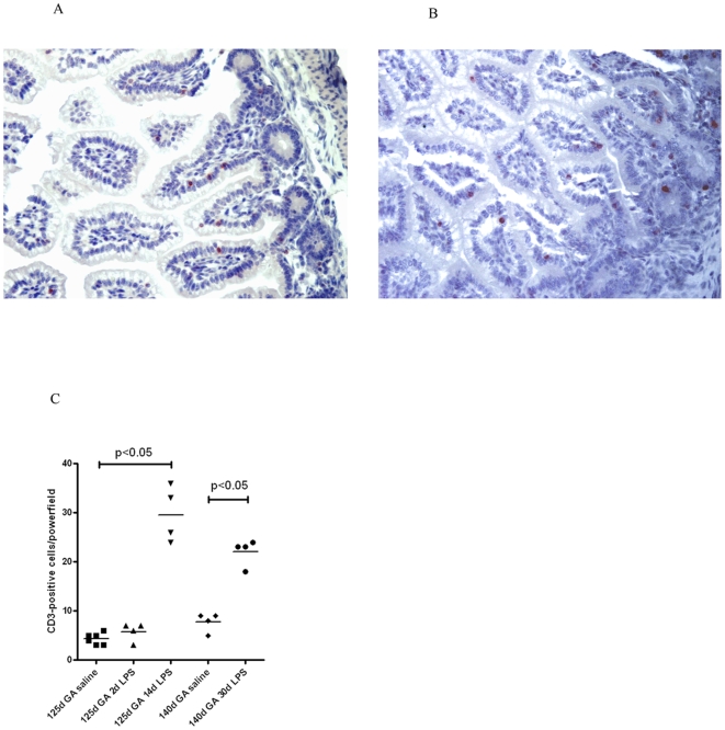
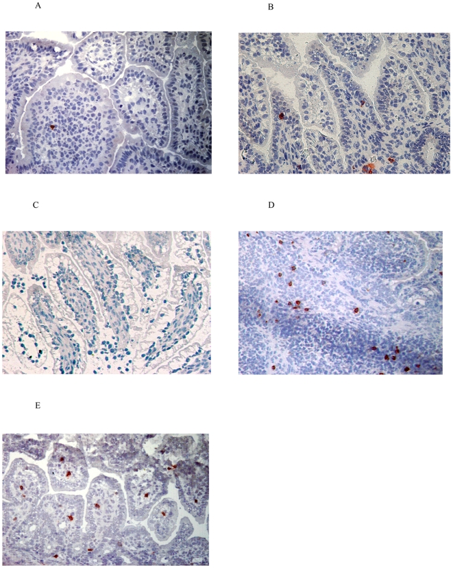
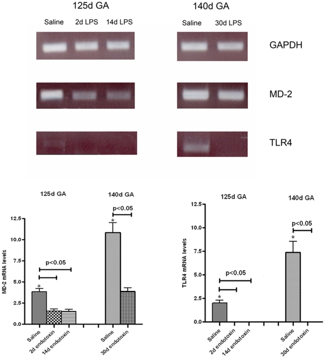
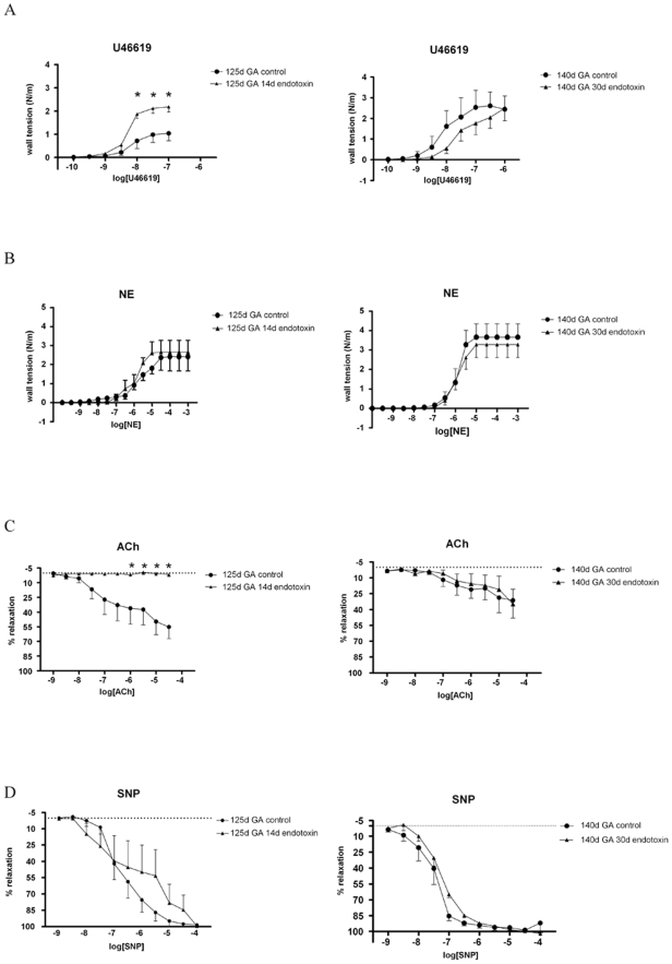
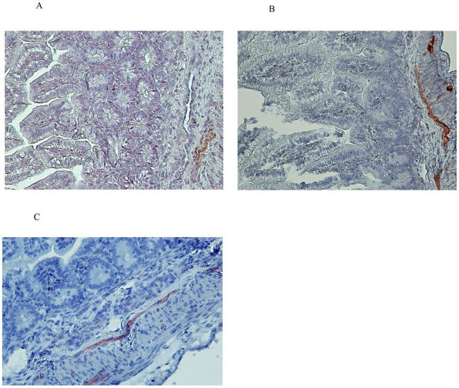
Chorioamnionitis is the most significant source of prenatal inflammation and preterm delivery. Prematurity and prenatal inflammation are associated with compromised postnatal developmental outcomes, of the intestinal immune defence, gut barrier function and the vascular system. We developed a sheep model to study how the antenatal development of the gut was affected by gestation and/or by endotoxin induced chorioamnionitis.Chorioamnionitis was induced at different gestational ages (GA). Animals were sacrificed at low GA after 2d or 14d exposure to chorioamnionitis. Long term effects of 30d exposure to chorioamnionitis were studied in near term animals after induction of chorioamnionitis. The cellular distribution of tight junction protein ZO-1 was shown to be underdeveloped at low GA whereas endotoxin induced chorioamnionitis prevented the maturation of tight junctions during later gestation. Endotoxin induced chorioamnionitis did not induce an early (2d) inflammatory response in the gut in preterm animals. However, 14d after endotoxin administration preterm animals had increased numbers of T-lymphocytes, myeloperoxidase-positive cells and gammadelta T-cells which lasted till 30d after induction of chorioamnionitis in then near term animals. At early GA, low intestinal TLR-4 and MD-2 mRNA levels were detected which were further down regulated during endotoxin-induced chorioamnionitis. Predisposition to organ injury by ischemia was assessed by the vascular function of third-generation mesenteric arteries. Endotoxin-exposed animals of low GA had increased contractile response to the thromboxane A2 mimetic U46619 and reduced endothelium-dependent relaxation in responses to acetylcholine. The administration of a nitric oxide (NO) donor completely restored endothelial dysfunction suggesting reduced NO bioavailability which was not due to low expression of endothelial nitric oxide synthase.Our results indicate that the distribution of the tight junctional protein ZO-1, the immune defence and vascular function are immature at low GA and are further compromised by endotoxin-induced chorioamnionitis. This study suggests that both prematurity and inflammation in utero disturb fetal gut development, potentially predisposing to postnatal intestinal pathology.


Conflict of interest statement

Competing Interests: The authors have declared that no competing interests exist.

Figures

Figure 1
Figure 1. Experimental design.
Gestational development and effects of chorioamnionitis were studied at two different gestational ages. At 125d GA, fetuses were comparable to 27 weeks of human gestation. Lambs were almost near term at 140d GA since term gestation is 147d. Chorioamnionitis was induced by a single injection of endotoxin under ultrasound guidance at 111d GA and at 123d GA. Animals were delivered at 125d GA and animals of the control group underwent the same procedure with an injection of saline. Animals of 140d GA had a separate control group to assess gestational changes.
Figure 2
Figure 2. Immunolocalisation of ZO-1 (red) in the fetal intestine.
At 125d GA, a fragmented staining pattern for ZO-1 was seen in control animals (A) or lambs exposed to endotoxin for 2 (B) or 14d (C). At 140d GA, a normal ZO-1 distribution was detected in control animals (D) that became disrupted upon endotoxin exposure for 30d (E). Magnification 200x. For inset, 1000x magnification was used.
Figure 3
Figure 3. At 14 and 30d after intraamniotic endotoxin injection, massive influx of PMNs was detected (A+B).
At 140d GA, some MPO positive cells were detected in control sections (C). For each experimental group mean cell counts of MPO positive cells are depicted per high-power field (D).
Figure 4
Figure 4. Intraamniotic endotoxin results in increased ileal T-cell density.
Significant increase of CD-3 immunoreactivity was seen at 14 and 30d after endotoxin exposure (A+B). For each experimental group mean cell counts of CD-3 positive cells are depicted per high-power field (C).
Figure 5
Figure 5. Endotoxin induced chorioamnionitis results in migration of gammadelta T-cells to sites of mucosal damage at late GA.
After 2d or 14d of endotoxin exposure, hardly any gammadelta T-cells were detected in the upper mucosa of the preterm intestine (A+B). At 140d GA, gammadelta T-cells were not detected in the lamina propria whereas lymphoid follicles in the basal mucosa became populated with gamma delta T-cells (C+D). 30d after endotoxin injection, increased numbers of gammadelta T-cells were found within areas of mucosal damage (E).
Figure 6
Figure 6. Ileal TLR4 and MD-2 mRNA was evaluated by ovine specific RT-PCR amplification of cDNA samples from healthy control lambs and lambs subjected to endotoxin induced chorioamnionitis.
Representative DNA fragments are shown for each group (n = 4). cDNAs were standardized for GAPDH content. Quantitative data were obtained by densitometric evaluation of RT-PCR products which were compared to a standard curve obtained by amplification of a serial dilution of highly concentrated cDNA. At 125d GA, TLR4 and MD-2 mRNA levels were significantly (*) lower compared to animals at 140d GA. TLR4 and MD-2 mRNA expression was significantly reduced following intraamniotic endotoxin injection for 2, 14 and 30d.
Figure 7
Figure 7. Thromboxane A2 mimic U46619 induced contractions of jejunal arteries increased with gestational age and were significantly (*) (P<0.05) augmented after intra-amniotic endotoxin injection in the 125d (P<0.05) but not in the 140d GA animals
(A). Concentration-dependent contractile effects of norepinephrine tend to increase (p>0.05) during gestation but remained unaltered during chorioamnionitis (B). Concentration-dependent relaxation of mesenteric arteries induced by acetylcholine was unaltered during gestation and was significantly (*) inhibited at 14d but not after 30d of endotoxin exposure (C). The relaxation response curve for sodium nitroprusside remained identical during gestation or chorioamnionitis (D).
Figure 8
Figure 8. Intraamniotic endotoxin induces eNOS expression.
Weak eNOS staining was seen at 2d post endotoxin treatment (A) while expression increased in animals exposed to endotoxin for 14 or days 30d (B-C).

References

    1. Goldenberg RL, Hauth JC, Andrews WW. Intrauterine infection and pretermdelivery. N Engl J Med. 2000;342:1500–1507. - PubMed
    1. Saji F, Samejima Y, Kamiura S, Sawai K, Shimoya K, et al. Cytokine production in chorioamnionitis. J Reprod Immunol. 2000;47:185–196. - PubMed
    1. Kramer BW, Kramer S, Ikegami M, Jobe AH. Injury, inflammation, and remodeling in fetal sheep lung after intra-amniotic endotoxin. Am J Physiol Lung Cell Mol Physiol. 2002;283:L452–459. - PubMed
    1. Kallapur SG, Jobe AH. Contribution of inflammation to lung injury and development. Arch Dis Child Fetal Neonatal Ed. 2006;91:F132–135. - PMC - PubMed
    1. Kramer BW, Moss TJ, Willet KE, Newnham JP, Sly PD, et al. Dose and time response after intraamniotic endotoxin in preterm lambs. Am J Respir Crit Care Med. 2001;164:982–988. - PubMed