. 2009 Sep;238(9):2235-53.
doi: 10.1002/dvdy.21989.

Genome-wide expression profiling in the Drosophila eye reveals unexpected repression of notch signaling by the JAK/STAT pathway

Affiliations

Genome-wide expression profiling in the Drosophila eye reveals unexpected repression of notch signaling by the JAK/STAT pathway

Maria Sol Flaherty et al. Dev Dyn. 2009 Sep.

Abstract

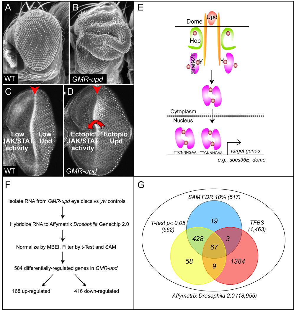
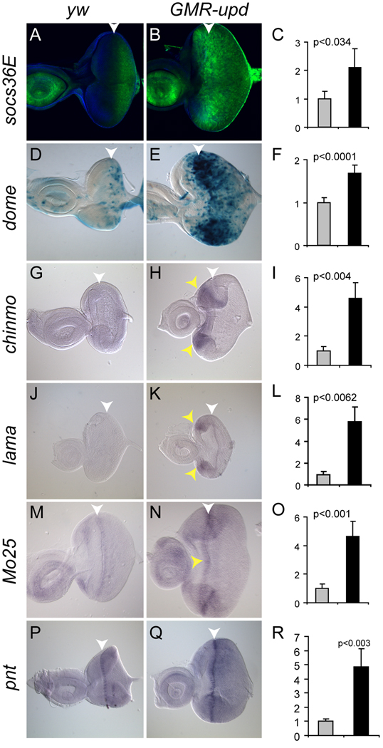
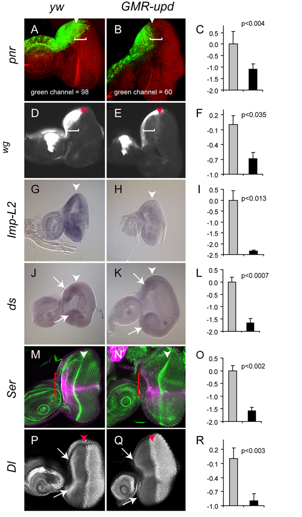
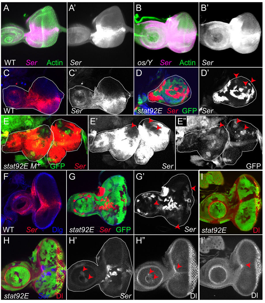
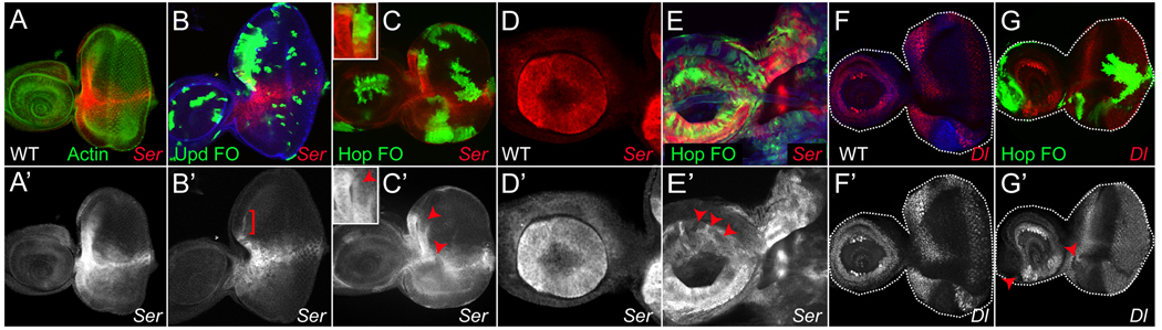
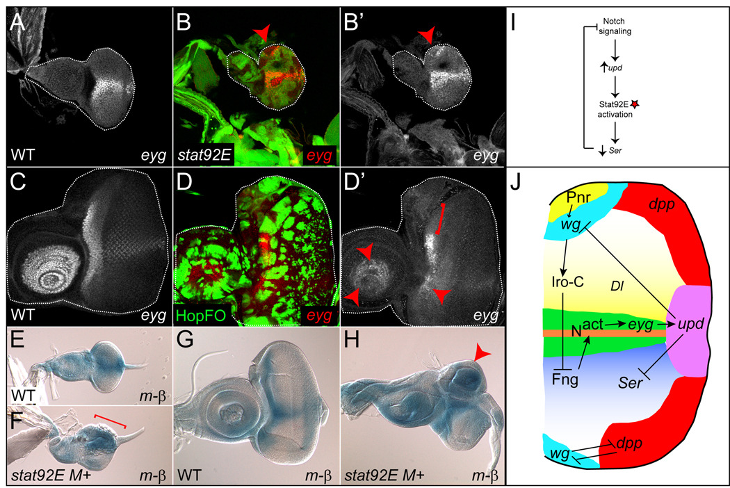
Although the JAK/STAT pathway regulates numerous processes in vertebrates and invertebrates through modulating transcription, its functionally relevant transcriptional targets remain largely unknown. With one jak and one stat (stat92E), Drosophila provides a powerful system for finding new JAK/STAT target genes. Genome-wide expression profiling on eye discs in which Stat92E is hyperactivated, revealed 584 differentially regulated genes, including known targets domeless, socs36E, and wingless. Other differentially regulated genes (chinmo, lama, Mo25, Imp-L2, Serrate, Delta) were validated and may represent new Stat92E targets. Genetic experiments revealed that Stat92E cell-autonomously represses Serrate, which encodes a Notch ligand. Loss of Stat92E led to de-repression of Serrate in the dorsal eye, resulting in ectopic Notch signaling and aberrant eye growth there. Thus, our micro-array documents a new Stat92E target gene and a previously unidentified inhibitory action of Stat92E on Notch signaling. These data suggest that this study will be a useful resource for the identification of additional Stat92E targets.


Figures

Figure 1
Figure 1. Over-expression of upd driven by the GMR promoter generates an enlarged-eye phenotype
(A,B) Scanning electron micrographs (100x). Wild type (WT) adult eye (A). GMR-upd adult eye (B). (C,D) Immuno-fluorescence image of a WT (C) or GMR-upd (D) third instar eye imaginal disc dissected at 110 hours AED and stained with phalloidin. In wild type third instar eye discs, JAK/STAT signaling is low or not detectable because the ligand Upd stops being synthesized in posterior midline cells at the onset of third instar (Bach et al., 2003; Ekas et al., 2006; Bach et al., 2007). In contrast, in GMR-upd eye discs, Upd is ectopically produced throughout third instar by cells posterior to the morphogenetic furrow. This ectopic Upd diffuses anteriorly and induces activation of the JAK/STAT pathway in undifferentiated anterior cells (D, arrow). (E) The Drosophila JAK/STAT pathway. Upd is a secreted cytokine that activates a transmembrane cytokine receptor Dome, which in turn activates the JAK tyrosine kinase Hop and the STAT transcription factor Stat92E. Activated Stat92E dimers translocate from the cytoplasm to the nucleus, where they induce growth by promoting the transcription of targets, only a few of which like dome and socs36E have been identified. (F) Micro-array design and outcome (see also text for details). Biological quintuples of individual eye discs were analyzed by Affymetrix Drosophila 2.0 GeneChip arrays to identify sets of mRNAs significantly modulated between yw and GMR-upd samples. (G) The statistical filtering of array data (SAM or T-test) was performed as described in Materials and Methods, and overlap with a set of genes harboring Stat92E binding sites (TFBS) is shown.
Figure 2
Figure 2. Up-regulated genes anterior to the furrow in GMR-upd discs
The expression of all genes was analyzed in mid-third instar eye discs. The morphogenetic furrow (MF) is marked by a white arrowhead. The Y-axis in the bar graphs represents relative mRNA abundance. (A,B) socs36E expression as assessed by the 10xSTAT92E-GFP transcriptional reporter (green) in yw control discs (A). socs36E is up-regulated anterior to the MF in GMR-upd discs (B). (C) socs36E is increased 2.10 fold in the GMR-upd micro-array. (D,E) dome-Gal4, UAS-lacZ (abbreviated dome) expression (blue) in control eye discs (D). dome is up-regulated in cells anterior to the MF in GMR-upd discs (E). (F) dome is increased 1.68 fold in the GMR-upd micro-array. (G,H) chinmo-RC (labeled chinmo) mRNA, as assessed by in situ hybridization, is absent in control discs (G) and is increased in cells anterior to the MF in GMR-upd discs (H, yellow arrowheads). (I) chinmo is increased 4.6 fold in the GMR-upd micro-array. (J,K) lama mRNA, as assessed by in situ hybridization, is not present in wild type discs (J). However, it is up-regulated in cells anterior to the MF in GMR-upd discs (K, yellow arrowheads). (L) lama is increased 5.44 fold in the GMR-upd micro-array. (M,N) Mo25 mRNA, as assessed by in situ hybridization, is present faintly in cells within and immediately surrounding the furrow in wild type discs (M). In GMR-upd discs Mo25 has a broader expression domain as well as increased expression levels (N, yellow arrowheads). (O) Mo25 is increased 4.65 fold in the GMR-upd micro-array. (P,Q) pnt mRNA, as assessed by in situ hybridization, is present in cells within the furrow in wild type discs (P). In GMR-upd discs, pnt is expressed at a significantly higher level in cells within the furrow, as well as at higher levels in cells adjacent to the furrow (Q). (R) pnt is increased 4.8 fold in the GMR-upd micro-array. Gray bar is yw and black bar is GMR-upd (C,F,I,L,O,R).
Figure 3
Figure 3. Down-regulated genes in GMR-upd
The expression of all genes was analyzed in mid-third instar eye discs. The morphogenetic furrow (MF) is marked by an arrowhead. The Y-axis in the bar graphs represents relative mRNA abundance. (A,B) pnr-Gal4, UAS-gfp (abbreviated pnr) (green) is expressed in dorsal peripodial cells in yw control discs (A, bracket). In GMR-upd discs, pnr is repressed in dorsal peripodial cells located “above” cells anterior to the MF (B, bracket). The mean area of pnr expression in wild type eye discs (A) is 98 pixel sq, while in GMR-upd (B) it is reduced to 60 pixels sq. (C) pnr is down-regulated 2.13 fold in the GMR-upd micro-array. (D,E) wg-lacZ (white) is expressed in cells anterior to the furrow at the lateral margins of the dorsal and ventral poles in control discs (D, bracket). In GMR-upd discs, wg expression is decreased in cells anterior to the MF (E, bracket). (F) wg is down-regulated 1.61 fold in the GMR-upd micro-array. (G,H) Imp-L2 mRNA, as assessed by in situ hybridization, is down-regulated in cells anterior to the MF in GMR-upd discs (H) as compared to yw controls (G). (I) Imp-L2 is decreased 5.08 fold in the GMR-upd micro-array. (J,K) ds is expressed in cells at the dorsal and ventral poles in controls discs (J, arrows). Its expression at the poles is greatly reduced in GMR-upd discs (K, arrows). (L) ds is decreased 3.14 fold in the GMR-upd micro-array. (M,N) Ser-lacZ (magenta) is expressed at the D–V boundary and in cells at the anterior edge of control discs (M, bracket). Ser is repressed in cells anterior to the MF in GMR-upd discs (N, bracket). (O) Ser is down-regulated 2.98 fold in the GMR-upd micro-array. (P,Q) In a wild type third instar eye disc, Dl-lacZ (white) is expressed in cells at the anterior margin of the disc and in cone cells posterior to the furrow (P, arrowheads). In GMR-upd discs, Dl is repressed in cells anterior to the MF (Q, arrow). Dl is decreased 1.86 fold in the GMR-upd micro-array (R). The brackets in A,B,D,E,M,N indicate the region in which gene expression is repressed in GMR-upd discs. Gray bar is yw and black bar is GMR-upd (C,F,I,L,O,R).
Figure 4
Figure 4. JAK/STAT signaling negatively regulates Ser and Dl
(A–B) Heterozygous os/+; Ser-LacZ/+ third instar eye discs show an expression pattern of Ser (magenta) that is indistinguishable from wild type (A,A’, compare Fig. 4A to Fig. 3M). In contrast, hemizygous os/Y; Ser-LacZ:+ third instar eye discs show ectopic expression of Ser in both the eye and antennal disc (B,B’). (C–G) Ser is ectopically expressed in stat92E clones. In a wild type second instar eye disc, Ser (red) is expressed in the ventral domain (C). Ser is ectopically expressed in mosaic stat92E clones, except those residing at the dorsal pole (D,D’, arrowheads). Large stat92E clones generated in a Minute background (labeled stat92E M+) lack expression of GFP. In stat92E M+ clones, Ser is ectopically expressed throughout the entire mutant tissue (E, red and E’, white), except in GFP+ heterozygous cells which contain a wild type copy of the stat92E gene (E, green, E’, arrowheads and E” white and arrowheads). In a wild type third instar disc, Ser (red) is expressed in cells at the D–V boundary and at the anterior lateral margin; it is excluded from the distal antenna (F). Ser (red) is ectopically expressed in stat92E clones residing in both the dorsal and ventral domains of the eye as well as those in the distal antenna (G,G’ arrowheads). (H–I) Ser (blue) and Dl (red) are ectopically expressed in stat92E clones. Dl is ectopically expressed in stat92E clones residing at the anterior margin of the eye disc (H,H”,I,I’, arrowheads in eye disc). In wild type disc, Dl is expressed in ring around A3 (see Suppl Fig 5A). However, in stat92E clones residing in the distal antenna, Dl expression is altered such that it now appears as a “dot” (H”, arrowhead in antennal disc) within the middle of ectopic Ser (H’, arrowheads in antennal disc). Single channel for Ser (A’-E’,G’,H’). Single channel for Dl (H”,I”). Phalloidin is green in A. Dlg is blue in C,D,F. Ser-LacZ was detected by anti-β-gal. stat92E397 clones lack GFP in D,E,G-I.
Figure 5
Figure 5. Activated Stat92E represses Ser
(A) Expression of Ser-LacZ (red) in a mid-third instar eye disc. (B) A large upd-expressing clone (green) located at the dorsal anterior margin of the eye significantly represses endogenous expression of Ser there (B’, bracket). (C) hop-expressing clones located at the dorsal anterior margin (magnified in inset), D–V midline or in the ventral domain significantly repress Ser (red) in a cell-autonomous manner (C’, arrowhead and inset). (D) Ser (red) is expressed in a ring around the distal antenna in a wild type eye-antennal disc. (E) hop-expressing clones repress Ser in a cell-autonomous manner in the antenna (E’, arrowheads). (F) Expression of Dl-lacZ in a wild type third instar eye-antennal disc. (G) a hop-expressing clone represses Dl-lacZ expression at the ventral anterior lateral margin in a cell-autonomous manner (G’, arrowhead in eye disc). Note the re-patterning induced by the hop-expressing clone in the antenna (G’, arrowhead in antennal disc). Single channel for Ser (A’-E’) and for Dl (F’,G”). Phalloidin is green in A. Dlg is blue in B,E. Ser-LacZ and Dl-LacZ were detected by anti-β-gal. upd- or hop-expressing clones are GFP+ in B,C,E,G.
Figure 6
Figure 6. Notch signaling is ectopically activated when JAK/STAT signaling is reduced
(A) eyg-lacZ (white) is expressed at the D–V midline in a wildtype (WT) late second instar eye disc. (B) eyg-LacZ (red) is ectopically expressed within a stat92E mosaic clone at the dorsal pole of the a second instar eye disc (B,B’, arrowhead). (C) In WT third instar, eyg-lacZ (white) is expressed at the anterior margin of the eye disc and in the distal antenna. (D) hop-expressing clones (green) cause repression of eyg-lacZ in a cell autonomous manner (D’, arrowheads and bracket). (E) The m-β reporter is normally expressed at the D–V boundary in a WT second instar eye disc. (F) However, in a second instar disc containing large stat92E M+ clones, this reporter is ectopically expressed throughout the dorsal eye disc (F, bracket), precisely in the region where Ser is ectopically expressed (see Fig. 4E). (G) In a WT third instar eye disc, the m-β reporter is normally expressed at the D–V boundary and at the anterior margin. (H) However, in a third instar disc containing large stat92E M+ clones, a circular, independent growth domain is observed in the dorsal eye that contains high levels of m-β reporter activity (arrowhead). (I) Model of negative feedback loop between Notch and JAK/STAT pathways. Notch regulates upd autonomously in the eye disc. Upd then acts on neighboring cells to activate Stat92E (red star). Stat92E represses Ser expression in a cell-autonomous manner. This results in reduction in Notch ligand levels and a decrease in Notch receptor activity. (J) Model of negative feedback loop between Notch and JAK/STAT pathways in the context of the developing eye disc. See text for details. eyg-LacZ was detected by anti-β-gal. Single channel for eyg-LacZ is (A,B’,C,D’). hop-expressing clones are GFP+ (D). m-β was detected by Xgal staining.

Similar articles

Cited by

References

    1. Agaisse H, Perrimon N. The roles of JAK/STAT signaling in Drosophila immune responses. Immunol Rev. 2004;198:72–82. - PubMed
    1. Agaisse H, Petersen UM, Boutros M, Mathey-Prevot B, Perrimon N. Signaling Role of Hemocytes in Drosophila JAK/STAT-Dependent Response to Septic Injury. Dev Cell. 2003;5:441–450. - PubMed
    1. Arbouzova NI, Bach EA, Zeidler MP. Ken & barbie selectively regulates the expression of a subset of Jak/STAT pathway target genes. Curr Biol. 2006;16:80–88. - PubMed
    1. Arbouzova NI, Zeidler MP. JAK/STAT signalling in Drosophila: insights into conserved regulatory and cellular functions. Development. 2006;133:2605–2616. - PubMed
    1. Ayala-Camargo A, Ekas LA, Flaherty MS, Baeg GH, Bach EA. The JAK/STAT pathway regulates proximo-distal patterning in Drosophila. Dev Dyn. 2007;236:2721–2730. - PubMed

Publication types

MeSH terms