. 2009 Jul 14;48(27):6412-22.
doi: 10.1021/bi900474g.

The two active sites of Thermotoga maritima CheA dimers bind ATP with dramatically different affinities

Affiliations

The two active sites of Thermotoga maritima CheA dimers bind ATP with dramatically different affinities

Anna K Eaton et al. Biochemistry. .

Abstract

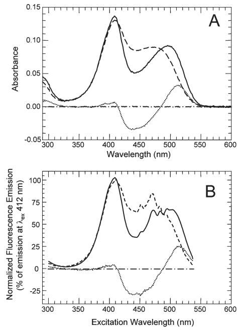
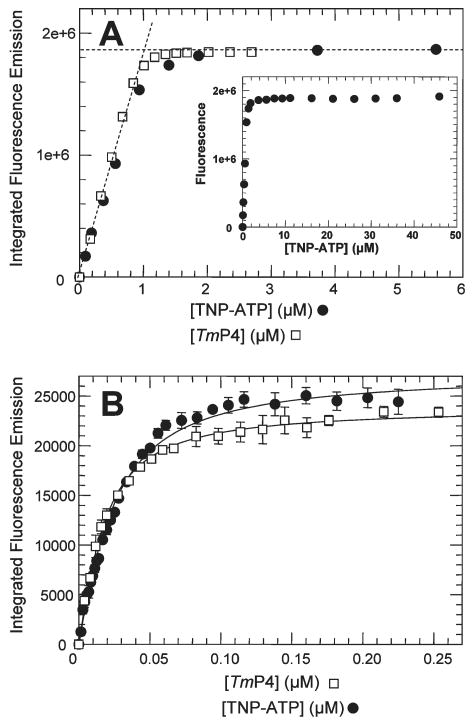
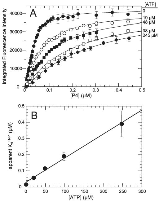
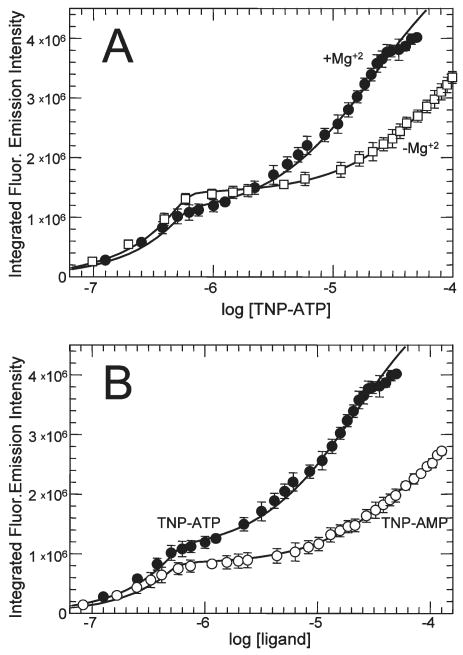
CheA is a central component of the chemotaxis signal transduction pathway that allows prokaryotic cells to control their movements in response to environmental cues. This dimeric protein histidine kinase autophosphorylates via an intersubunit phosphorylation reaction in which each protomer of the dimer binds ATP, at an active site located in its P4 domain and then catalyzes transfer of the gamma-phosphoryl group of ATP to the His(45) side chain within the P1 domain of the trans protomer. Here we utilize the fluorescent nucleotide analogue TNP-ATP [2'(3')-O-(2,4,6-trinitrophenyl)adenosine 5'-triphosphate] to investigate the two ATP-binding sites of the Thermotoga maritima CheA dimer (TmCheA) and the single site of the isolated TmP4 domain (a monomer). We define the affinity of CheA for TNP nucleotides and, by competition, for unmodified ATP. The two ATP-binding sites of the TmCheA dimer exhibit dramatically different affinities for TNP-ATP (K(d1)(TNP) approximately 0.0016 muM and K(d2)(TNP) approximately 22 muM at 4 degrees C in the presence of Mg(2+)) as well as for ATP (K(d1)(ATP) approximately 6 muM and K(d2)(ATP) approximately 5000 muM at 4 degrees C in the presence of Mg(2+)) and in their ability to influence the fluorescence of bound TNP-ATP. The ATP-binding site of the isolated TmP4 domain interacts with ATP and TNP-ATP in a manner similar to that of the high-affinity site of the TmCheA dimer. These results suggest that the two active sites of TmCheA homodimers exhibit large differences in their interactions with ATP. We consider possible implications of these differences for the CheA autophosphorylation mechanism and for CheA function in bacterial cells.


Figures

Figure 1
Figure 1
Schematic diagram of the functional organization of CheA and the mechanism of autophosphorylation. Domains P1–P5 of CheA from T. maritima, E. coli, and S. typhimurium have been investigated by a variety of high-resolution methods, including X-ray crystallography (1, 33, 39, 47, 63) and NMR methods (34, 35, 66), as well as by lower-resolution methods such as protease sensitivity (37). The numbers written below the top protomer in the diagram indicate the domain boundaries (amino acid numbers) of T. maritima CheA. The jagged lines linking P1 to P2 and P2 to P3 are intended to depict flexible linkers. The functional role of each domain is based on this structural information as well as on biochemical and genetic experiments (17, 37, 38, 57). As depicted, CheA autophosphorylation occurs via a trans intradimer mechanism in which the ATP bound to the P4 domain of one protomer provides the phosphoryl group that is used to phosphorylate His45 on the partner protomer.
Figure 2
Figure 2
Effect of TmP4 on the absorbance and fluorescence properties of TNP-ATP. (A) The absorbance spectrum of 4.9 μM TNP-ATP (in TnM buffer) was recorded in the absence (− − −) and presence (—) of 12 μM TmP4. Also shown is the difference spectrum (⋯) calculated by subtracting the TNP-ATP spectrum from the spectrum of TNP-ATP with TmP4. Spectra were recorded vs a baseline scan of TnM buffer; a buffer vs buffer scan (− · −) is shown. (B) Fluorescence excitation spectra were recorded (λem = 540 nm) for 4.9 μM TNP-ATP in the absence (− − −) and presence (—) of 12 μM TmP4; plotted spectra were normalized relative to the emission intensity observed for a λex of 412 nm. Also shown are the calculated difference spectrum (⋯) (TNP-ATP scan subtracted from TmP4/TNP-ATP scan) and a buffer–buffer baseline (− · −).
Figure 3
Figure 3
Effect of TmP4 on the fluorescence excitation and emission spectra of TNP-ATP. (A) The main panel shows the fluorescence excitation spectrum of 4.9 μM TNP-ATP (in TnM buffer) in the absence (---) and presence (—) of 12 μM TmP4 using an emission wavelength of 540 nm. The inset plots the signal-to-background ratio as a function of the excitation wavelength for this same sample set (arrows at 410 and 520 nm). (B) Fluorescence emission spectrum of 4.9 μM TNP-ATP (in TnM buffer) recorded in the absence (---) and presence (—) of 12 μM TmP4 using an excitation wavelength of 520 nm. Also shown is the corrected buffer emission scan (− · −).
Figure 4
Figure 4
Fluorescence-monitored binding of TNP-ATP to TmP4. (A) Results of titrations in which successive aliquots of TNP-ATP stock solutions were added to a 2.5 mL sample of TmP4 (1 μM) (●) or aliquots of TmP4 were added to a 2.5 mL sample of TNP-ATP (1 μM) (□). The intersection point of the dashed lines indicates a binding stoichiometry of 1:1. The inset shows the TNP-ATP titration results at higher concentrations. (B) Results of titrations at low protein and ligand concentrations. Successive aliquots of TNP-ATP stock solutions were added to a 2.5 mL sample of TmP4 (0.015 μM) (●), or aliquots of TmP4 were added to a 2.5 mL sample of TNP-ATP (0.013 μM) (□). The lines represent the best fits obtained using DynaFit to fit the data using a single-binding site model. This analysis indicated a Kd of 0.011 μM for the titration of TNP-ATP with TmP4 and a Kd of 0.016 μM for the titration of TmP4 with TNP-ATP. Error bars indicate standard deviations for three independent experiments.
Figure 5
Figure 5
Competition between ATP and TNP-ATP for the TmP4 binding site. (A) TmP4 aliquots were added to TNP-ATP samples (0.02 μM) containing 0 (●), 19 (○), 48 (■), 98 (□), and 245 μM ATP (◆). The solid lines are the best fits obtained using DynaFit to fit the data to a model of a single binding site; this analysis defined an apparent KdTNP value at each concentration of competitor (ATP). (B) Apparent dissociation constants from panel A plotted as a function of ATP concentration.
Figure 6
Figure 6
Fluorescence-monitored binding of TNP-ATP to TmCheA. (A) Results of titrations in which successive aliquots of TNP-ATP stock solutions were added to a 2.5 mL sample of TmCheA (0.6 μM dimer). The line represents the best fit of the data generated using DynaFit and a two-site binding model. This analysis gave the following values: Kd1TNP = 0.0029 μM, Kd2TNP = 22 μM, IMFCT.AA = 2.0 × 10⁶ cps/μM, and IMFCT.AA.T = 9.5 × 10⁶ cps/μM. The inset shows the same results plotted using a log scale for [TNP-ATP] to better depict the extremely biphasic nature of the binding curves. (B) Results of a reverse titration in which aliquots of a concentrated TmCheA solution were added to a 0.28 μM solution of TNP-ATP. The intersection point of the dashed lines indicates a binding stoichiometry of one TNP-ATP per CheA dimer and a value of 2.0 × 10⁶ cps/μM for IMFCT.AA.
Figure 7
Figure 7
Binding and ATP inhibition of binding of TNP-ATP to the high-affinity site of TmCheA. (A) Binding of TNP-ATP to TmCheA was monitored fluorometrically as successive aliquots of TNP-ATP were added to 0.003 μM TmCheA (dimer concentration) (●) and when aliquots of TmCheA were added to 0.002 μM TNP-ATP (□). The lines represent the best fits of the data generated using DynaFit and a one-site binding model. This analysis indicated a Kd1TNP of 0.0018 μM for the titration of CheA with excess TNP-ATP and a Kd1TNP of 0.0014 μM for the titration of TNP-ATP with excess TmCheA. Error bars indicate the standard deviations for three independent experiments. (B) Effect of competitor ATP on the apparent Kd1TNP of TmCheA.
Figure 8
Figure 8
Competition between ATP and TNP-ATP for TmCheA binding sites. (A) TNP-ATP aliquots were added to 0.6 μM TmCheA samples (dimer concentration) containing 0 (●), 2.34 (○), 5.9 (■), and 23.4 mM ATP (□). Binding was monitored by following fluorescence emission. Not shown (to avoid overcrowding) are titration results at 11.9 mM ATP. The solid lines are the best fits obtained using Dyna Fit to fit the data to a model in which each CheA dimer has two binding sites that make unequal contributions to the observed fluorescence change (2 × 10⁶ cps/μM for the first site and 7.5 × 10⁶ cps/μM for the second site, as defined in Figure 6). (B) Effect of competitor ATP on the apparent Kd2ATP (determined in panel A). Analysis of this relationship (as described in the legend of Figure 5) indicates a Kd2ATP of 5000 ± 1000 μM.
Figure 9
Figure 9
TmCheA binding to TNP-AMP and to TNP-ATP in the absence of Mg2+. (A) Aliquots of TNP-ATP were added to 0.6 μM TmCheA samples (dimer concentration) in buffer either containing 20 mM MgCl2 (●) or lacking MgCl2 (and containing 20 mM Na2EDTA) (□); the resulting fluorescence signals were monitored and analyzed as described in the legend of Figure 6. The lines represent the best fit of the data using DynaFit and a two-site binding model. This analysis indicated that in the absence of Mg2+, Kd1TNP = 0.003 μM, Kd2TNP = 100 μM, IMFCT.AA = 2.4 × 10⁶ cps/μM, and IMFCT.AA.T = 8.9 × 10⁶ cps/μM. In the presence of Mg2+, Kd1TNP = 0.0016 μM, Kd2TNP = 22 μM, IMFCT.AA = 2 × 10⁶ cps/μM, and IMFCT.AA.T = 9.5 × 10⁶ cps/μM. (B) Aliquots of TNP-ATP (●) or TNP-AMP (○) were added to 0.6 μM (dimer) TmCheA samples in buffer containing 20 mM MgCl2. Binding was assessed by monitoring fluorescence. The TNP-ATP titration data are the same as those presented in panel A. The lines represent the best fit of the data using DynaFit and a two-site binding model, indicating that for TNP-AMP binding Kd1TNP = 0.003 μM, Kd2TNP = 75 μM, IMFCT.AA = 1.4 × 10⁶ cps/μM, and IMFCT.AA.T = 6.3 × 10⁶ cps/μM.
Figure 10
Figure 10
Models for nucleotide binding to dimeric TmCheA. (A) Negative cooperativity model. (B) Asymmetric dimer model. Nucleotide is depicted as a black circle. CheA protomers are depicted as circles (high-affinity conformation) and squares (low-affinity conformation). Protomer colors indicate the ability of the protomer to enhance the fluorescence of bound TNP-ATP: light orange indicating a weaker ability and red indicating a stronger ability.

Similar articles

Cited by

References

    1. Bilwes A, Alex L, Crane BR, Simon MI. Structure of CheA, a signal-transducing histidine kinase. Cell. 1999;96:131–141. - PubMed
    1. Bilwes A, Quezada CM, Croal LR, Crane BR, Simon MI. Nucleotide binding by the histidine kinase CheA. Nat Struct Biol. 2001;8:353–360. - PubMed
    1. Bilwes AMP, Quezada CM, Simon M, Crane BR. Histidine Kinases in Signal Transduction. Academic Press; San Diego: 2003. pp. 48–74.
    1. Borkovich KA, Simon MI. The dynamics of protein phosphorylation in bacterial chemotaxis. Cell. 1990;63:1339–1348. - PubMed
    1. Bornhorst JA, Falke JJ. Attractant regulation of the aspartate receptor-kinase complex: Limited cooperative interactions between receptors and effects of the receptor modification state. Biochemistry. 2000;39:9486–9493. - PMC - PubMed

Publication types