. 2009 Aug 25;101(11):645-59.
doi: 10.1042/BC20090042.

Wnt5 is required for notochord cell intercalation in the ascidian Halocynthia roretzi

Affiliations

Wnt5 is required for notochord cell intercalation in the ascidian Halocynthia roretzi

Tomoko Niwano et al. Biol Cell. .

Abstract

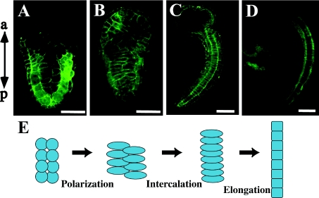
Background information: In the embryos of various animals, the body elongates after gastrulation by morphogenetic movements involving convergent extension. The Wnt/PCP (planar cell polarity) pathway plays roles in this process, particularly mediolateral polarization and intercalation of the embryonic cells. In ascidians, several factors in this pathway, including Wnt5, have been identified and found to be involved in the intercalation process of notochord cells.

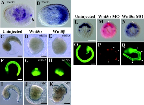
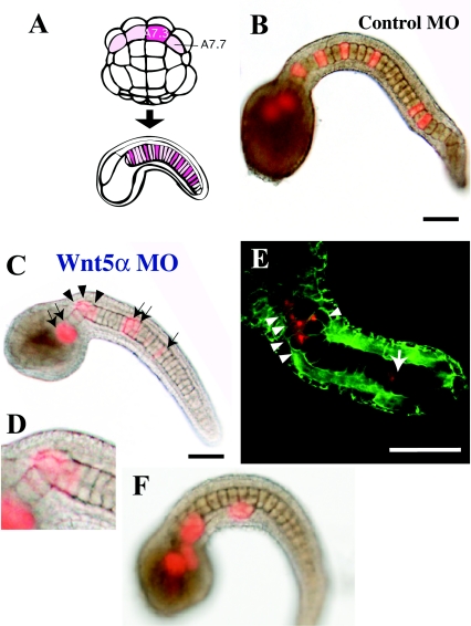
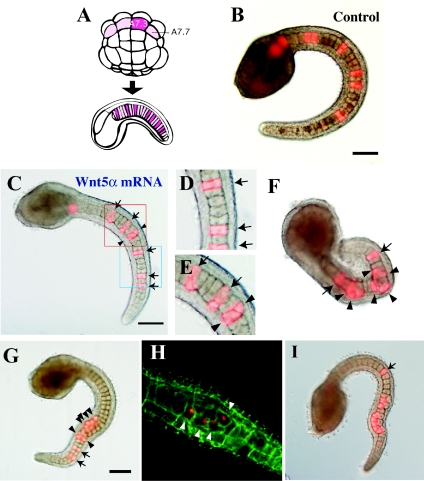
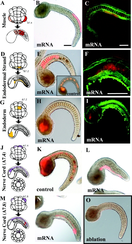
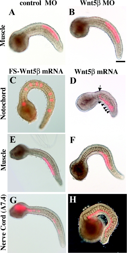
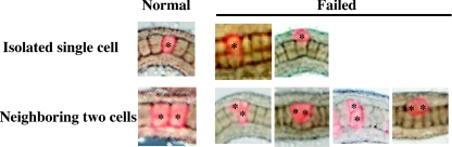
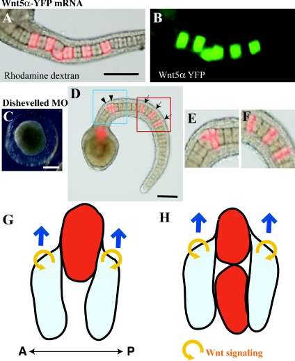
Results: In the present study, the role of the Wnt5 genes, Hr-Wnt5alpha (Halocynthia roretzi Wnt5alpha) and Hr-Wnt5beta, in convergent extension was investigated in the ascidian H. roretzi by injecting antisense oligonucleotides and mRNAs into single precursor blastomeres of various tissues, including notochord, at the 64-cell stage. Hr-Wnt5alpha is expressed in developing notochord and was essential for notochord morphogenesis. Precise quantitative control of its expression level was crucial for proper cell intercalation. Overexpression of Wnt5 proteins in notochord and other tissues that surround the notochord indicated that Wnt5alpha plays a role within the notochord, and is unlikely to be the source of polarizing cues arising outside the notochord. Detailed mosaic analysis of the behaviour of individual notochord cells overexpressing Wnt5alpha indicated that a Wnt5alpha-manipulated cell does not affect the behaviour of neighbouring notochord cells, suggesting that Wnt5alpha works in a cell-autonomous manner. This is further supported by comparison of the results of Wnt5alpha and Dsh (Dishevelled) knockdown experiments. In addition, our results suggest that the Wnt/PCP pathway is also involved in mediolateral intercalation of cells of the ventral row of the nerve cord (floor plate) and the endodermal strand.

Conclusion: The present study highlights the role of the Wnt5alpha signal in notochord convergent extension movements in ascidian embryos. Our results raise the novel possibility that Wnt5alpha functions in a cell-autonomous manner in activation of the Wnt/PCP pathway to polarize the protrusive activity that drives convergent extension.


Figures

Figure 1
Figure 1. Convergent extension movement of ascidian notochord
Confocal images of frontal section of H. roretzi stained with Alexa Fluor® 488 phalloidin to visualize cellular boundaries. (A) At the initial tail-bud stage, 40 post-mitotic notochord cells are aligned in two bilateral lines. Anterior is at the top. (B) Each notochord cell is polarized, elongates along the left–right axis and starts mediolateral intercalation. (C) The notochord is made of disc-shaped cells aligned in a single line after intercalation. (D) At the mid tail-bud stage, each notochord cell elongates in a process of post-convergent extension elongation. In consequence, the tail further elongates. (E) Representation of notochord morphogenesis. a, anterior; p, posterior. Scale bar, 100 μm.
Figure 2
Figure 2. Effects of Wnt5 MOs and mRNAs injected into eggs
(A, B) Expression of Hr-Wnt5α and Hr-Wnt5β genes at the neurula stage in developing notochord and muscle cells respectively. Anterior is to the left. Arrowhead indicates Hr-Wnt5α expression in the posterior pole, which is concentrated Hr-Wnt5α maternal mRNA, as it is a member of postplasmic/PEM RNAs in ascidians (Sasakura et al. 1998). (CE) Morphology of uninjected and Wnt5α and Wnt5β mRNA-injected embryos at the tail-bud stage. (FH) Detection of the notochord differentiation marker antigen, Not-1, in uninjected and Wnt5α and Wnt5β mRNA-injected embryos. (IK) Morphology of uninjected, Wnt5α and Wnt5β MO-injected embryos at the tail-bud stage respectively. (L) Expression of Bra gene in ten notochord precursor blastomeres at the 110-cell stage. (M) Embryo co-injected with Wnt5α MO and a lineage tracer (rhodamine dextran) into a single notochord precursor (A7.3) blastomere of the 64-cell embryo. At the 110-cell stage, two sister blastomeres are labelled with red fluorescence (white arrowheads). (N) The same embryo normally expressed the Bra gene in notochord precursors, including the labelled cells. (O) The Not-1 antigen is expressed in notochord. The embryo was slightly overstained with the antibody, and the signal is spread in the trunk region. (P) At the tail-bud stage, several descendants of the injected blastomere can be recognized from their content of red fluorescent label. (Q) The same embryo expressed Not-1 antigen in notochord cells including the injected and labelled cells (arrowheads). Scale bar, 100 μm.
Figure 3
Figure 3. Effects of Wnt5α MO injected into a notochord precursor blastomere of the 64-cell embryo
(A) Lineage illustration of notochord. Four pink blastomeres of the 64-cell embryo (vegetal view, anterior is up) give rise to the major part of the notochord in the tail. A7.3 and A7.7 are the notochord precursors. Each blastomere divides three times and gives rise to eight notochord cells randomly intercalated in the notochord. Injected blastomeres and their descendants are indicated in dark pink. (B) Tail-bud embryos co-injected with the lineage tracer and control universal MO into the A7.3 blastomere. The dark spot in the centre of each notochord cell is the nucleus. (C) Injection of Wnt5α MO. Arrows indicate normally intercalated notochord cells that are labelled with red fluorescence. Arrowheads show cells that failed to intercalate. (D) Closer view of the neck region of (C). (E) Another example of injection of Wnt5α MO observed with a confocal microscope. Cell boundaries are demarcated by green fluorescence. (F) Embryos that showed a severe phenotype. Labelled notochord cells are clustered and not incorporated into the aligned notochord. Scale bar, 100 μm.
Figure 4
Figure 4. Effects of Wnt5α mRNA injected into a notochord precursor blastomere
(A) Lineage illustration of notochord. (B) Tail-bud embryos co-injected with the lineage tracer and control FS Wnt5α mRNA into the A7.3 blastomere. (C) Injection of Wnt5α mRNA. Arrows indicate normally intercalated notochord cells. Arrowheads show cells that failed to intercalate. (D) Closer view of the blue rectangle in (C). Three notochord cells successfully intercalated. (E) Closer view of the red rectangle in (C). The two cells marked with arrowheads failed to intercalate with each other. (F–I) Four examples of Wnt5α mRNA-injected embryos to show the wide spectrum of abnormality in intercalation. In (I), post-convergent elongation of notochord cells is taking place. Scale bar, 100 μm.
Figure 5
Figure 5. Effects of Wnt5α mRNA injected into various tissue precursors
(A, D, G, J, M) Lineage illustration of muscle, endodermal strand, endoderm and nerve cord (A7.4 and A7.8) respectively (Nishida, 1987). Injected blastomeres and their descendants are indicated in dark colours. (B, E, H, K, L, N) Bright-field images are merged with fluorescent images to show the position of labelled descendant cells. (C, F, I) Confocal views. Cell boundaries are demarcated by green fluorescence. (B, C) The muscle precursor was injected. (E, F) The precursor of the endodermal strand was injected. The notochord is normal, but intercalation of the endodermal strand, which is present on this side of the notochord, was prevented as the labelled cells did not intercalate with the cells of the other bilateral side as they formed a continuous line. (F) is a confocal closer view of such a specimen. (E, insert) Control embryo. The endodermal strand is intermittently labelled in the tail. (H, I) The endoderm precursor was injected. (K, L) Control mRNA and Wnt5α mRNA were injected into the precursor of the ventral row (floor plate) of the nerve cord respectively. In the control, the nerve cord is intermittently labelled in the tail, indicating intercalation of left and right descendants. In Wnt5α mRNA-injected embryos, the labelled cells failed to intercalate as they formed a continuous line. (N) Wnt5α mRNA was injected into the precursor of the lateral row of the nerve cord in addition to two muscle cells at the tip of the tail (red). (O) Every A-line nerve cord precursor blastomere was ablated at the 64-cell stage by bursting them with injected seawater. The notochord of the embryo developed normally, indicating that the neural tube is dispensable for notochord morphogenesis. Scale bar, 100 μm.
Figure 6
Figure 6. Effects of Wnt5β MO and mRNA injected into various tissue precursors
(A, B) Control and Wnt5β MO were injected into the muscle precursor (B7.4) blastomere as Wnt5β is expressed in muscle. (CH) Control FS Wnt5β mRNA and wild-type Wnt5β mRNA were injected into the precursors of notochord (A7.3), muscle (B7.4) and the ventral row of nerve cord (A7.4). Arrow indicates a normally intercalated notochord cell. Arrowheads show cells that failed to intercalate. (G) In the control, the nerve cord is intermittently labelled in the tail, indicating intercalation of left and right descendants. (H) In Wnt5β mRNA-injected embryos, the labelled cells failed to intercalate as they formed a continuous line. Scale bar, 100 μm.
Figure 7
Figure 7. Mosaic analysis of Wnt knockdown and overexpression within the notochord
A single notochord precursor blastomere was injected with Wnt5α MO or mRNA, or with Dsh MO. Examples of normal and abnormal intercalation of the labelled cells (asterisks) in the two situations where labelled cells are present in isolation or two labelled cells are adjacent. Abnormal positioning of labelled cells is categorized into two and four types respectively. The detailed frequency of each abnormality is shown is Table 2.
Figure 8
Figure 8. Wnt5α may function in a cell-autonomous manner
(A, B) The lineage tracer (rhodamine dextran; red) and mRNA encoding the Wnt5α–YFP fusion protein (green) were co-injected into a notochord precursor blastomere. The green signal remains close to the injected descendant cells. (C) An embryo in which Dsh MO was injected into fertilized eggs. (D) An embryo in which Dsh MO was injected into a notochord precursor blastomere. Arrows indicate normally intercalated notochord cells. Arrowheads show cells that failed to intercalate. (E, F) Closer views of the blue and red rectangles shown in (D). Scale bar, 100 μm. (G) A descendant cell of the MO- and mRNA-injected blastomere (red) is present in isolation. Flanking normal cells (light blue) may migrate on the surface of the anomalous cell. Blue arrows indicate the direction of movement of the normal cells. Yellow circular arrows refer to the autocrine Wnt5α action. Anterior is to the left. A, anterior; P, posterior. (H) When anomalous cells are closely adjacent they fail to migrate on each other. See the text for further details.

References

    1. Akanuma T., Hori S., Darras S., Nishida H. Notch signaling is involved in neurogenesis in the ascidian embryos. Dev. Genes Evol. 2002;212:459–472. - PubMed
    1. Barrow J.R. Wnt/PCP signaling: a veritable polar star in establishing patterns of polarity in embryonic tissues. Semin. Cell Dev. Biol. 2006;17:185–193. - PubMed
    1. Christian J.L. BMP, Wnt and Hedgehog signals: how far can they go? Curr. Opin. Cell Biol. 2000;2:244–249. - PubMed
    1. Cloney R.A. Development of the ascidian notochord. Acta Embryol. Morphol. Exp. 1964;7:111–130.
    1. Fanto M., McNeill H. Planar polarity from flies to vertebrates. J. Cell Sci. 2004;117:527–533. - PubMed

Publication types

LinkOut - more resources