Review
. 2009 Jun 12;421(1):1-15.
doi: 10.1042/BJ20081016.

At the crossroads of homoeostasis and disease: roles of the PACS proteins in membrane traffic and apoptosis

Affiliations
Review

At the crossroads of homoeostasis and disease: roles of the PACS proteins in membrane traffic and apoptosis

Robert T Youker et al. Biochem J. .

Abstract

The endomembrane system in mammalian cells has evolved over the past two billion years from a simple endocytic pathway in a single-celled primordial ancestor to complex networks supporting multicellular structures that form metazoan tissue and organ systems. The increased organellar complexity of metazoan cells requires additional trafficking machinery absent in yeast or other unicellular organisms to maintain organ homoeostasis and to process the signals that control proliferation, differentiation or the execution of cell death programmes. The PACS (phosphofurin acidic cluster sorting) proteins are one such family of multifunctional membrane traffic regulators that mediate organ homoeostasis and have important roles in diverse pathologies and disease states. This review summarizes our current knowledge of the PACS proteins, including their structure and regulation in cargo binding, their genetics, their roles in secretory and endocytic pathway traffic, interorganellar communication and how cell-death signals reprogramme the PACS proteins to regulate apoptosis. We also summarize our current understanding of how PACS genes are dysregulated in cancer and how viral pathogens ranging from HIV-1 to herpesviruses have evolved to usurp the PACS sorting machinery to promote virus assembly, viral spread and immunoevasion.


Figures
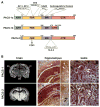
Figure 1
Figure 1. Domain organization of the PACS proteins
(A) Schematic of PACS-1 and PACS-2 illustrating the proposed domains and residues important for partner protein binding. (B) In situ hybridization of PACS-1 and PACS-2. Left-hand panels, coronal sections of rat brain stained with PACS-1 or PACS-2 cRNA probes. DG, dentate gyrus; Hc, hippocampus; Ctx, cortex; Th, thalamus; MM, medial mammillary nucleus. Middle panels, darkfield staining of hippocampus showing neuronal and glial labelling. (*), alignment marker; white arrows, neurons; black arrows, glia. Right-hand panels, PACS-1 and PACS-2 in consecutive serial sections of the testis. Arrows mark seminiferous tubules with inverse staining of PACS-1 and PACS-2. Sense probes showed no staining.
Figure 2
Figure 2. Phylogenetic analysis of PACS genes
Non-redundant protein sequences of the PACS family members were obtained from the SwissProt and NCBI databases (Accession numbers used: A0NG63, Q5ZJW8, Q95QA3, Q7YRF1, P36225, Q8MRS5, Q4PGG5, Q13367, Q6VY07, Q86VP3, Q7Z6E9, A4HVI7, Q17EX1, Q5PSV9, Q8K212, Q3V3Q7, Q6J6J0, Q4SQP4, Q9PVV4, Q9HGI6, A5PKS9). The protein sequences were aligned using ClustalW and were manually examined/modified for their accuracy within the non-conserved domains that flank conserved domains. The protein alignments were used to obtain nucleotide alignments, using a PERL script, as described previously [172]. The tree topology was estimated from the DNA alignment using the DNAML module from the PHYLIP package and our data set was analysed in PAML using appropriate models (M8, M8a) [172]. A LRT (likelihood ratio test) was used to search for positive selection. Positive selection (Ω > 1) was tested by applying an empirical Bayesian approach which allows for all types of selection (purifying, neutral and positive), including nesting of models which do not allow positive selection [–175]. The statistical significance of our tree was estimated through a LRT test on a null tree (M8a), which does not allow for positive selection [176]. Consistent with studies on other protein families, trees generated using different programs (DNAPARS and DNADIST) do not affect the analysis. A. aegypti, Aedes aegypti; A. gambiae, Anopheles gambiae; AP3β1, AP-3 complex subunit β-1; AP3β2, AP-3 complex subunit β-2; BRCA1, breast cancer type 1 susceptibility protein homologue; B. taurus, Bos taurus; C. familiaris, Canis familiaris; D. hansenii, Debaryomyces hansenii; D. rerio, Danio rerio; DTL, denticleless protein homologue; ERF2, eukaryotic peptide chain release factor GTP-binding subunit; G. gallus, Gallus gallus; L. infantum, Leishmania infantum; H. sapiens, Homo sapiens; MAP4, microtubule-associated protein 4; MDC1, mediator of DNA damage checkpoint protein 1; M. musculus, Mus musculus; P. pygmaeus, Pipistrellus pygmaeus; RBBP6, retinoblastoma-binding protein 6R. norvegicus, Rattus norvegicus; T. nigroviridis, Tetraodon nigroviridis; U. maydis, Ustilago maydis.
Figure 3
Figure 3. Positively selected residues of PACS-1 and PACS-2
Multiple-sequence alignment of conserved regions in PACS-1 and PACS-2 based on the phylogenetic analysis of the PACS proteins (Figure 2). For simplicity, eight PACS-1 and two PACS-2 sequences were used to generate the alignment. Residues shaded in black are > 80 % conserved. Residues shaded dark grey ~ 60–65 % conserved and residues shaded in light grey are ~ 50 % conserved. Residues highlighted in yellow are positively selected, which was determined by measuring the rate of non-synonymous to synonymous substitutions (Ω), which is an unequivocal indicator of evolution [172]. Residues with a value Ω = 1 are under neutral selection whereas residues with Ω values < 1 or > 1 suggest that the residues are under a purifying or positive selection respectively [174]. Of the residues, 2.7 % in PACS-1 exhibit strong positive selection (Ω > 1). Residues were shaded using genedoc. Secondary structure prediction (PsiPredict) for PACS-1 (above alignment) and PACS-2 (below alignment) was colour-coded the same as the constructs in Figure 1(A). Predicted regions of disorder (PONDR) for PACS-1 and PACS-2 are stippled on the schematic of the secondary structure. A. gambiae, Anopheles gambiae; D. rerio, Danio rerio; G. gallus; Gallus gallus; H. sapiens, Homo sapiens; M. musculus, Mus musculus; T. nigroviridis, Tetraodon nigroviridis.
Figure 4
Figure 4. Ab initio model of PACS-1 FBR
Ab initio modelling of the PACS-1 FBR (residues 117–300) was performed using Rosetta++ [177] and the predicted structure was energy minimized using Insight II (Accelrys Insight II modelling software). The tertiary structure is represented as a ribbon diagram surrounded by a semi-transparent surface projection. Acidic residues are coloured red, basic residues are coloured blue, residues required for adaptor binding are coloured orange, residues required for GGA binding are coloured yellow and residues positively selected through evolution are coloured pink. Left-hand panel, top view looking down on the major groove. Right-hand panel, the image is rotated 90° towards the viewer. Images were created using PyMOL (DeLano Scientific; http://pymol.sourceforge.net/). An interactive three-dimensional version of this Figure is available at http://www.BiochemJ.org/bj/421/0001/bj4210001add.htm.
Figure 5
Figure 5. Working model of PACS-1/GGA3-regulated trafficking of CI-MPR
GGA3 transports phosphorylated CI-MPR from the TGN to endosomes [180]. At the endosome, PACS-1 binds to the VHS domain of GGA3 through its FBR and recruits CK2 to phosphorylate Ser388 on GGA3 and phosphorylate Ser278 on PACS-1, thus releasing the autoregulatory domain and activating PACS-1 for cargo binding. CK2 may also phosphorylate additional sites on GGA3 [181] to promote release from endosomal membranes. Phosphorylated GGA3 dissociates, and activated PACS-1 binds to CI-MPR. Bound PACS-1 then recruits AP-1 to transport CI-MPR back to the TGN. It is not known whether CK2 bound to PACS-1 can phosphorylate the CI-MPR tail or other cargo molecules. An animated version of this Figure is available at http://www.BiochemJ.org/bj/421/0001/bj4210001add.htm.
Figure 6
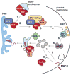
Figure 6. ‘Signalling’ model of Nef-mediated down-regulation of cell-surface MHC-I
Step 1: Nef binds PACS-2 through its acidic cluster (EEEE65) and is targeted to the late Golgi/TGN. Step 2: at the TGN, Nef PXXP75 binds and activates a TGN-localized SFK. Step 3: the activated Nef–SFK complex recruits and activates ZAP-70/Syk. Tyrosine-phosphorylated ZAP-70/Syk then binds a class I PI3K. Step 4: Nef-stimulated class I PI3K generates PIP3 on the inner leaflet of the plasma membrane. Step 5: an ARF6 guanine-nucleotide-exchange factor (ARF6-GEF) is recruited to PIP3 at the plasma membrane. Step 6: the recruited ARF6-GEF in turn activates ARF6. Step 7: MHC-I is rapidly endocytosed from the plasma membrane to internal endosomal compartments. Steps 8 and 9: sequestration of newly internalized MHC-I molecules to a paranuclear region requires the Nef Met20 and its interaction with AP-1. The precise step of Nef-mediated MHC-I down-regulation requiring PACS-1 is unclear but the ability of PACS-1 to bind Nef and AP-1 raises the possibility that PACS-1 may contribute to the Met20-dependent internalization of MHC-I molecules.
Figure 7
Figure 7. Working model of Akt/14-3-3/PP2A regulation of PACS-2 apoptotic activity
Under non-stressed conditions, apoptotic PACS-2 is phosphorylated on Ser437 and held inactive in the cytosol through binding to 14-3-3 isoforms. Loss of Akt signalling or apoptotic activation of PP2A leads to dephosphorylation of PACS-2 and release from 14-3-3. ‘Activated’ PACS-2 binds and facilitates translocation of full-length Bid to mitochondria, inducing cytochrome c release. This pathway parallels the previously described 14-3-3-regulated translocation of the pro-apoptotic protein Bad [182,183].

References

    1. Lee MC, Miller EA, Goldberg J, Orci L, Schekman R. Bi-directional protein transport between the ER and Golgi. Annu Rev Cell Dev Biol. 2004;20:87–123. - PubMed
    1. McMahon HT, Mills IG. COP and clathrin-coated vesicle budding: different pathways, common approaches. Curr Opin Cell Biol. 2004;16:379–391. - PubMed
    1. Palade G. Intracellular aspects of the process of protein synthesis. Science. 1975;189:347–358. - PubMed
    1. Fewell SW, Travers KJ, Weissman JS, Brodsky JL. The action of molecular chaperones in the early secretory pathway. Annu Rev Genet. 2001;35:149–191. - PubMed
    1. Gu F, Crump CM, Thomas G. Trans-Golgi network sorting. Cell Mol Life Sci. 2001;58:1067–1084. - PMC - PubMed

Publication types

Substances