Expression of Ebolavirus glycoprotein on the target cells enhances viral entry
- PMID: 19505320
- PMCID: PMC2699336
- DOI: 10.1186/1743-422X-6-75
Expression of Ebolavirus glycoprotein on the target cells enhances viral entry
Abstract
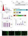
Background: Entry of Ebolavirus to the target cells is mediated by the viral glycoprotein GP. The native GP exists as a homotrimer on the virions and contains two subunits, a surface subunit (GP1) that is involved in receptor binding and a transmembrane subunit (GP2) that mediates the virus-host membrane fusion. Previously we showed that over-expression of GP on the target cells blocks GP-mediated viral entry, which is mostly likely due to receptor interference by GP1.
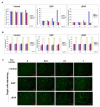
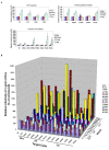
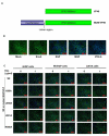
Results: In this study, using a tetracycline inducible system, we report that low levels of GP expression on the target cells, instead of interfering, specifically enhance GP mediated viral entry. Detailed mapping analysis strongly suggests that the fusion subunit GP2 is primarily responsible for this novel phenomenon, here referred to as trans enhancement.
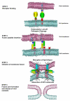
Conclusion: Our data suggests that GP2 mediated trans enhancement of virus fusion occurs via a mechanism analogous to eukaryotic membrane fusion processes involving specific trans oligomerization and cooperative interaction of fusion mediators. These findings have important implications in our current understanding of virus entry and superinfection interference.
Figures
Similar articles
-
Biological Characterization of Conserved Residues within the Cytoplasmic Tail of the Pichinde Arenaviral Glycoprotein Subunit 2 (GP2).J Virol. 2019 Oct 29;93(22):e01277-19. doi: 10.1128/JVI.01277-19. Print 2019 Nov 15. J Virol. 2019. PMID: 31462569 Free PMC article.
-
Spontaneous Mutation at Amino Acid 544 of the Ebola Virus Glycoprotein Potentiates Virus Entry and Selection in Tissue Culture.J Virol. 2017 Jul 12;91(15):e00392-17. doi: 10.1128/JVI.00392-17. Print 2017 Aug 1. J Virol. 2017. PMID: 28539437 Free PMC article.
-
Ebola virus glycoprotein needs an additional trigger, beyond proteolytic priming for membrane fusion.PLoS Negl Trop Dis. 2011 Nov;5(11):e1395. doi: 10.1371/journal.pntd.0001395. Epub 2011 Nov 15. PLoS Negl Trop Dis. 2011. PMID: 22102923 Free PMC article.
-
Filovirus entry: a novelty in the viral fusion world.Viruses. 2012 Feb;4(2):258-75. doi: 10.3390/v4020258. Epub 2012 Feb 7. Viruses. 2012. PMID: 22470835 Free PMC article. Review.
-
Structure-function relationship of the mammarenavirus envelope glycoprotein.Virol Sin. 2016 Oct;31(5):380-394. doi: 10.1007/s12250-016-3815-4. Epub 2016 Aug 4. Virol Sin. 2016. PMID: 27562602 Free PMC article. Review.
Cited by
-
The cytoplasmic domain of Marburg virus GP modulates early steps of viral infection.J Virol. 2011 Aug;85(16):8188-96. doi: 10.1128/JVI.00453-11. Epub 2011 Jun 15. J Virol. 2011. PMID: 21680524 Free PMC article.
-
An Ebola Virus-Like Particle-Based Reporter System Enables Evaluation of Antiviral Drugs In Vivo under Non-Biosafety Level 4 Conditions.J Virol. 2016 Sep 12;90(19):8720-8. doi: 10.1128/JVI.01239-16. Print 2016 Oct 1. J Virol. 2016. PMID: 27440895 Free PMC article.
-
Chemically Modified Human Serum Albumin Potently Blocks Entry of Ebola Pseudoviruses and Viruslike Particles.Antimicrob Agents Chemother. 2017 Mar 24;61(4):e02168-16. doi: 10.1128/AAC.02168-16. Print 2017 Apr. Antimicrob Agents Chemother. 2017. PMID: 28167539 Free PMC article.
-
Expression of an immunogenic Ebola immune complex in Nicotiana benthamiana.Plant Biotechnol J. 2011 Sep;9(7):807-16. doi: 10.1111/j.1467-7652.2011.00593.x. Epub 2011 Feb 1. Plant Biotechnol J. 2011. PMID: 21281425 Free PMC article.
-
Optimization of 4-Aminopiperidines as Inhibitors of Influenza A Viral Entry That Are Synergistic with Oseltamivir.J Med Chem. 2020 Mar 26;63(6):3120-3130. doi: 10.1021/acs.jmedchem.9b01900. Epub 2020 Mar 2. J Med Chem. 2020. PMID: 32069052 Free PMC article.
References
Publication types
MeSH terms
Substances
Grants and funding
LinkOut - more resources
Full Text Sources
Other Literature Sources
Medical

