. 2009 Sep 29;163(1):213-21.
doi: 10.1016/j.neuroscience.2009.05.073. Epub 2009 Jun 6.

Schwann cell influence on motor neuron regeneration accuracy

Affiliations

Schwann cell influence on motor neuron regeneration accuracy

R D Madison et al. Neuroscience. .

Abstract

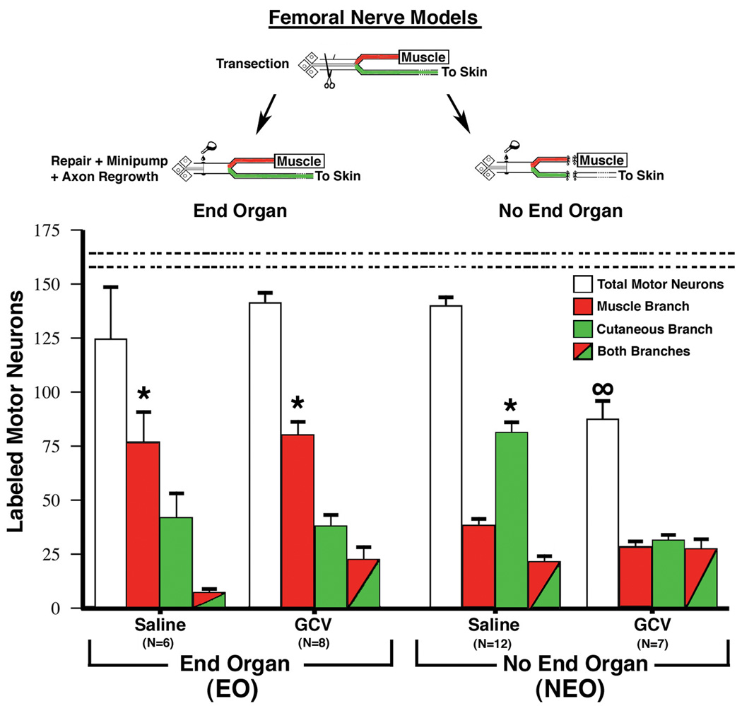
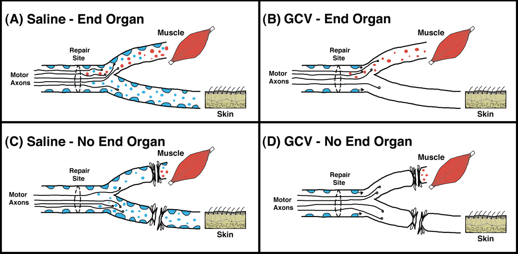
Extensive peripheral nerve injuries can result in the effective paralysis of the entire limb or distal portions of the limb. The major determinant of functional recovery after lesions in the peripheral nervous system is the accurate regeneration of axons to their original target end-organs. We used the mouse femoral nerve as a model to study motor neuron regeneration accuracy in terms of regenerating motor neurons projecting to their original terminal pathway to quadriceps muscle vs. the inappropriate pathway to skin. Using a variety of surgical manipulations and the selective removal of Schwann cells in the distal nerve via molecular targeting, we have examined the respective roles of end-organ influence (muscle) vs. Schwann cells in this model system. We found evidence of a hierarchy of trophic support that regulates motor neuron regeneration accuracy with muscle contact being the most potent, followed by the number or density of Schwann cells in the distal nerve branches. Manipulating the relative levels of these sources of influence resulted in predictable projection patterns of motor neurons into the terminal pathway either to skin or to muscle.


Figures

Figure 1
Figure 1
Representative high-power views of 1µm plastic-embedded sections stained with toluidine blue. WT and transgenic TK animals received a crush lesion of the sciatic nerve and implantation of a minipump to deliver ganciclovir (GCV; 20 mg/kg/day) for the first seven days after surgery. Nerve segments distal to the crush lesion were harvested 7, 14, or 35 days post-injury. Panels are from the 14-day time point. A) WT animal. Note the numerous Schwann cell nuclei (identified by their characteristic dense staining and elongated or irregular morphology; arrowheads). A few white blood cells are also present (identified as larger moderately dark cells with rounded or oblong cell bodies and nuclei; arrows). B) Transgenic animal (TK). Compared to the WT animal note the paucity of Schwann cells but similar density of white blood cells. Size bar in A represents 10 microns for both panels. The quantification of such sections is shown in figure 2.
Figure 2
Figure 2
Counts of the number of Schwann cells (A) and white blood cells (B) in 1µm plastic-embedded sections in distal nerve at various times post-injury following continuous GCV administration during the first 7-days. Data expressed as mean ± SEM per mm2, N=4 TK animals and N=4 WT animals at each time point. A, top panel: There is a rapid and dramatic increase in the number of Schwann cells in WT animals compared to uninjured controls by 2–3 weeks, representing a greater than 15-fold. A similar increase (11-fold) in TK animals is not seen until 5 weeks post-injury and represents the ingrowth of Schwann cells from the proximal nerve that accompany regenerating axons. *=p<.05 between WT and TK. B, bottom panel: In contrast to the dramatic differences in the number of Schwann cells between WT and TG animals, there are no differences in the number of inflammatory cells (white blood cells). Compared to uninjured controls, both experimental groups show significant increases in the number of white blood cells typical of Wallerian degeneration.
Figure 3
Figure 3
Quantitative assessment of regeneration accuracy of motor neurons obtained 8 weeks after femoral nerve repair in adult transgenic TK mice. In the End-Organ model (EO) both terminal branches remained intact to their respective end-organs of muscle and skin. In the No End-Organ model (NEO) all end-organ contact was prevented by transecting, ligating, and capping both terminal branches equidistant from the parent nerve bifurcation. Eight weeks after femoral nerve repair application of labeled dextran tracers to the two terminal nerve branches just distal to the bifurcation of the parent nerve allowed quantification of the number of motor neurons projecting solely to one of the branches (red or green alone) or simultaneously to both branches (both colors). In the unlesioned femoral nerve motor neurons are only retrogradely labeled from the muscle branch. In the EO model significantly more motor neurons were retrogradely labeled from the muscle branch compared to the cutaneous branch when saline was delivered for the first seven days after surgery. The same result was found when ganciclovir (GCV) was delivered, which selectively removed the original population of Schwann cells distal to the nerve lesion (see text for details). Very different results were obtained in the NEO model. When saline was delivered significantly more motor neurons projected to the cutaneous branch. When GCV was delivered about 1/3 of motor neurons projected solely to the muscle branch, 1/3 solely to the cutaneous branch, and 1/3 simultaneously to both branches. In addition, the total number of motor neurons that regenerated was significantly less than the number of motor neurons retrogradely labeled from control nerves (dashed horizontal lines, 161±4). Data is expressed as the mean ± SEM, *= p<.05 between the two branches, ∞ = p<.05 total motor neurons compared to control.
Figure 4
Figure 4
Model of motor neuron regeneration accuracy in the femoral nerve. We posit two main sources of trophic support for regenerating motor neurons and their axons; Schwann cells and muscle. Schwann cells in both terminal nerve branches are represented as blue, and trophic support from Schwann cells is represented as small blue circles. Trophic support originating from muscle is represented by small red circles. (A) When trophic support from both Schwann cells and muscle is present, more motor neurons project to the muscle branch and reinnervate muscle, indicating the potent influence of muscle on such regeneration. (B) When the original population of Schwann cells distal to the nerve lesion is removed via targeting with ganciclovir (GCV), the trophic support from muscle is still intact and once again more motor neurons project to the muscle branch and reinnervate muscle. Since GCV is only delivered during the first seven days, Schwann cells that grow into the distal nerve along with the regenerating axons after this time period will not be eliminated. (C) When trophic support from end-organs is prevented, but the original population of Schwann cells is left intact, more motor neurons project to the cutaneous branch because it has more Schwann cells and thus more trophic support from Schwann cells. This suggests that in the absence of the overriding support from muscle, it is the number and density of Schwann cells that determines the motor neuron projection choice. (D) When end-organ influences are prevented and Schwann cells distal to the nerve lesion are removed via targeting with ganciclovir (GCV), fewer motor neurons regenerate overall and those that do show no preference for either of the terminal nerve branches.

Similar articles

Cited by

References

    1. Abercrombie M. Estimation of nuclear population from microtome sections. Anat. Rec. 1946;4:239–246. - PubMed
    1. Al-Majed AA, Neumann CM, et al. Brief electrical stimulation promotes the speed and accuracy of motor axonal regeneration. J. Neurosci. 2000;20(7):2602–2608. - PMC - PubMed
    1. Bignami A, Eng LF, et al. Localization of the glial acidic protein in astrocytes by immunofluorescence. Brain Res. 1972;43:429–435. - PubMed
    1. Borrelli E, Heyman R, et al. Targetting of an inducible toxic phenotype in animal cells. Proc. Natl. Acad. Sci. USA. 1988;85:7572–7576. - PMC - PubMed
    1. Brewster ME, Raghavan K, et al. Enhanced delivery of ganciclovir to the brain through the use of redox targeting. Antimicrob. Agents Chemother. 1994;38:817–823. - PMC - PubMed

Publication types

MeSH terms