Review
. 2009 Aug;19(4):261-9.
doi: 10.1016/j.semcancer.2009.02.013. Epub 2009 Feb 24.

JC virus: an oncogenic virus in animals and humans?

Affiliations
Review

JC virus: an oncogenic virus in animals and humans?

Melissa S Maginnis et al. Semin Cancer Biol. 2009 Aug.

Abstract

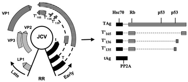
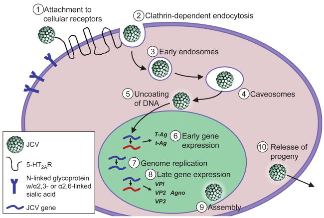
JC virus (JCV) is a human polyomavirus of the Polyomaviridae family, which also includes BK virus and simian vacuolating virus 40 (SV40). JC virus was first isolated in 1971 from the brain of a patient with Progressive Multifocal Leukoencephalopathy (PML). Like other polyomaviruses, JCV has a restricted host range. The virus infects the majority of the human population with seroconversion occurring during adolescence. JCV has a limited and specific tissue tropism infecting the kidney and oligodendrocytes and astrocytes in the central nervous system (CNS). Initial JCV infection is generally asymptomatic in immunocompetent hosts, and it establishes a persistent infection in the kidney and possibly bone marrow. In immunocompromised individuals JCV can cause a lytic infection in the CNS and lead to development of the fatal, demyelinating disease PML. The name polyoma is derived from the Greek terms: poly, meaning many, and oma, meaning tumors, owing to the capacity of this group of viruses to cause tumors. JCV inoculation of small animal models and non-human primates, which are not permissive to a productive JCV infection, leads to tumor formation. Given the ubiquitous nature of the virus and its strong association with cancer in animal models, it is hypothesized that JCV plays a role in human cancers. However, the role for JCV in human cancers and tumor formation is not clear. Some researchers have reported an association of JCV with human cancers including brain tumors, colorectal cancers, and cancers of the gastrointestinal tract, while other groups report no correlation. Here, we review the role of JCV in cancers in animal models and present the findings on JCV in human cancers.


Conflict of interest statement

Conflict of Interest Statement

The authors declare that they have no conflicts of interest.

Figures

Fig. 1
Fig. 1
Molecular map of the JCV genome. The JCV genome is a circular, dsDNA genome, approximately 5.1 kb in size. The viral genome is divided into the early coding region and the late coding region, which are separated by the non-coding control region or regulatory region (RR). Transcription of viral genes is temporally regulated and occurs in a bidirectional manner as depicted by the black arrows. Early genes include small t antigen, large T antigen, T′(135), T′ (136), and T′ (165). Late genes include LP1, VP1, VP2, and VP3. The JCV t- and T- antigen and T′ proteins and host-cellular protein interaction domains are shown on the right. Specifically, heat-shock 70 (Hsc70), retinoblastoma (Rb), p53, and PP2A are indicated due to their role in cellular transformation.
Fig. 2
Fig. 2
The JCV life cycle. JCV infection of host cells is initiated by attachment to cellular receptors, α-2,3- or α-2,6-linked sialic acid and serotonin receptor 5-HT2AR. The virus is internalized into cells by clathrin-dependent endocytosis. JCV then traffics through early endosomes and caveosomes to the nucleus. In the nucleus viral early gene transcription occurs, followed by viral DNA replication and late gene transcription. After production of the viral structural proteins VP1, VP2, and VP3, progeny virions are assembled in the nucleus and released.
Fig. 3
Fig. 3
Glioblastoma in an owl monkey brain. Shown is a representative image of a glioblastoma induced in the right cerebral hemisphere (bottom panel) in the brain of an owl monkey after inoculation with tumor suspensions from an explanted JCV-induced owl monkey astrocytoma. The top panel shows the left cerebral hemisphere free of tumors. The juvenile owl monkey was inoculated intracerebrally with suspensions of JCV-infected primary human fetal glial cells and developed a glioblastoma 28 months later. JCV DNA was recovered from tumor cells and demonstrated homology to JCV Mad-1 yet had a 19-base pair deletion in one of the 98-base pair repeats of the regulatory region. The viral sequence showed similarity to the JCV neurooncogenic strain, Mad-4. This new virus was named JCV-586. Image was kindly provided by Eugene Major.

References

    1. Padgett BL, Walker DL, ZuRhein GM, Eckroade RJ, Dessel BH. Cultivation of papova-like virus from human brain with progressive multifocal leucoencephalopathy. Lancet. 1971;1(7712):1257–60. - PubMed
    1. Major EO, Amemiya K, Tornatore CS, Houff SA, Berger JR. Pathogenesis and molecular biology of progressive multifocal leukoencephalopathy, the JC virus-induced demyelinating disease of the human brain. Clin Microbiol Rev. 1992;5(1):49–73. - PMC - PubMed
    1. Knowles WA, et al. Population-based study of antibody to the human polyomaviruses BKV and JCV and the simian polyomavirus SV40. J Med Virol. 2003;71(1):115–23. - PubMed
    1. Bofill-Mas S, Girones R. Excretion and transmission of JCV in human populations. J Neurovirol. 2001;7(4):345–9. - PubMed
    1. Bofill-Mas S, Pina S, Girones R. Documenting the epidemiologic patterns of polyomaviruses in human populations by studying their presence in urban sewage. Appl Environ Microbiol. 2000;66(1):238–45. - PMC - PubMed

Publication types

MeSH terms