

Reduction of the total injection volume of contrast material with a short injection duration in 64-detector row CT coronary angiography
- PMID: 19505968
- PMCID: PMC3487251
- DOI: 10.1259/bjr/23203690
Reduction of the total injection volume of contrast material with a short injection duration in 64-detector row CT coronary angiography
Abstract
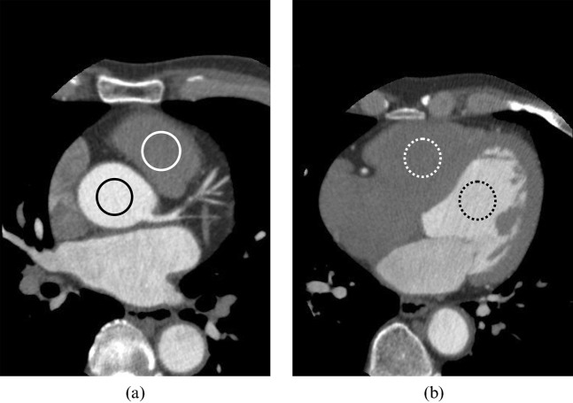
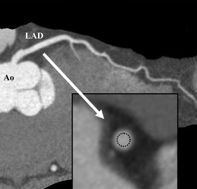
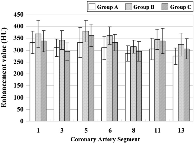
The ability of short injection duration of contrast material to reduce the total injection volume in 64-detector row CT coronary angiography was investigated. 45 patients were divided into three groups: (i) those receiving 0.8 ml kg(-1) of contrast material (350 mgI ml(-1)) injected with a fixed duration of 14 s (Group A; n = 16); (ii) those receiving 0.8 ml kg(-1) of contrast material injected with a fixed duration of 10 s (Group B; n = 15); and (iii) those receiving 0.7 ml kg(-1) of contrast material injected with a fixed duration of 10 s (Group C; n = 14). All patients then received 20 ml of saline. Contrast densities of the ascending aorta and proximal and distal coronary arteries were assessed where vessel diameters were >2.0 mm. The mean enhancement value in the ascending aorta for Group B was significantly higher than that for Groups A and C (p<0.05), whereas there was no significant difference between Groups A and C. All enhancement values in the coronary arteries were higher than 250 Hounsfield units. The mean enhancement value for each coronary artery in Group B was significantly higher than that for Group A (p<0.05), whereas there was no significant difference between Groups A and C. In conclusion, a short injection duration allows a reduction in the total volume of contrast material from 0.8 ml kg(-1) to 0.7 ml kg(-1) while a steady contrast enhancement is maintained in the ascending aorta and coronary arteries.
Figures
References
-
- Achenbach S, Giesler T, Ropers D, Ulzheimer S, Derlien H, Schulte C, et al. Detection of coronary artery stenoses by contrast-enhanced, retrospectively electrocardiographically-gated, multislice spiral computed tomography. Circulation 2001;103:2535–8 - PubMed
-
- Nieman K, Oudkerk M, Rensing BJ, van Ooijen P, Munne A, van Geuns RJ, et al. Coronary angiography with multi-slice computed tomography. Lancet 2001;357:599–603 - PubMed
-
- Bley TA, Ghanem NA, Foell D, Uhl M, Geibel A, Bode C, et al. Computed tomography coronary angiography with 370-millisecond gantry rotation time: evaluation of the best image reconstruction interval. J Comput Assist Tomogr 2005;29:1–5 - PubMed
-
- Nikolaou K, Flohr T, Knez A, Rist C, Wintersperger B, Johnson T, et al. Advances in cardiac CT imaging: 64-slice scanner. Int Journal Cardiovascular Imaging 2004;20:535–40 - PubMed
Publication types
MeSH terms
Substances
LinkOut - more resources
Full Text Sources
Other Literature Sources
