

doi: 10.1128/AAC.01623-08.
Epub 2009 Jun 8.
Nukacin ISK-1, a bacteriostatic lantibiotic
Affiliations
- PMID: 19506061
- PMCID: PMC2715603
- DOI: 10.1128/AAC.01623-08
Item in Clipboard
Nukacin ISK-1, a bacteriostatic lantibiotic
Antimicrob Agents Chemother.
2009 Aug.
Abstract
We determined the mode of action of nukacin ISK-1. It did not cause membrane potential dissipation or the efflux of ATP or K(+) ions from the cells of a sensitive bacterial strain; however, it blocked the membrane depolarization activity of nisin. Nukacin ISK-1-treated cells had single arrangements of cells without the formation of a complete septum. A remarkable reduction in cell wall width was also observed, but cytoplasmic content was unaffected. We concluded that nukacin ISK-1 is bacteriostatic.
Figures
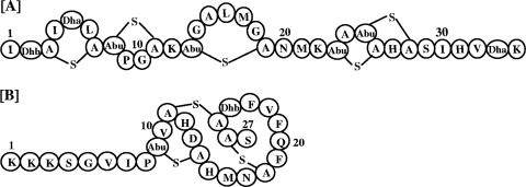
Structure of nisin A (A) and nukacin ISK-1 (B). A-S-A, lanthionine; Abu-S-A, 3-methyllanthionine; Abu, aminobutyrate; Dha, dehydroalanine; Dhb, dehydrobutyrine.
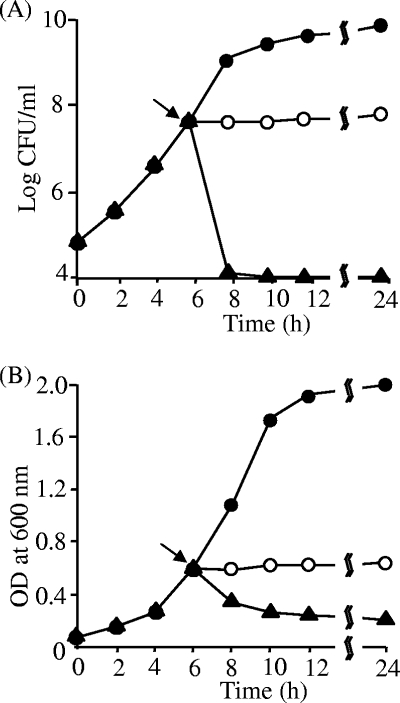
Bacteriostatic or bactericidal nature of nukacin ISK-1. CFU were counted (A) and optical density (OD) determined (B). The indicator bacterial strain L. sakei JCM 1157T was grown to the mid-log phase, and the culture was divided into three aliquots. Control cells (•) were not treated, and cells in the other two aliquots were treated with either nukacin ISK-1 (○) or nisin (▴). The lantibiotics (10× MIC) were added, the turbidity (OD) measured, and the CFU determined by the spread plate method after plating the cells on MRS agar in 10-fold serial dilutions. The arrow indicates the point at which nukacin ISK-1/nisin was added. This experiment was performed twice with high reproducibility. Representative data were used for the figure.
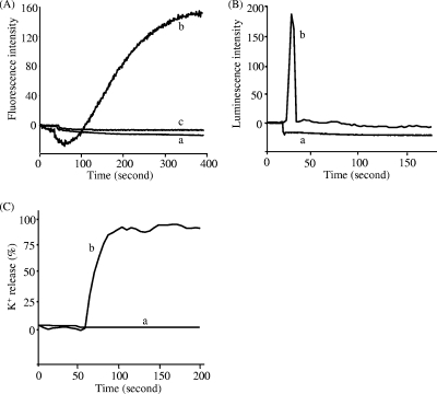
Antibacterial action of nukacin ISK-1. (A) Dissipation of the cell membrane potential by nukacin ISK-1 in normal cells (a), nisin A in normal cells (b), and nisin A in nukacin ISK-1-treated cells (c). The dissipation was examined by observing the fluorescence emitted by DiS-C3 (3,3′-dipropylthiadicarbocyanine iodide). (B) Efflux of ATP molecules from nukacin ISK-1-treated cells (a) and nisin A-treated cells (b), detected by using a Lucifer-HS ATP detection kit. (C) Efflux of K+ ions from nukacin ISK-1-treated cells (a) and nisin A-treated cells (b), detected by using potassium-binding benzofuran isophthalate-AM (PBFI). K+ efflux is expressed in relation to the total amount of K+ released after the addition of 1 μM nisin (100% value). These experiments were carried out with the lantibiotics at 10× MIC, using B. subtilis JCM 1465T as an indicator. These experiments were performed twice with high reproducibility. Representative data were used for the figure.
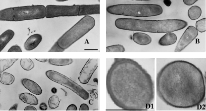
Overview of transmission electron micrographs of B. subtilis JCM 1465T cells. (A) The control cells were not treated with any lantibiotics. (B and C) Micrographs of cells that were treated with nukacin ISK-1 (B) and nisin A (C) at 10× MIC for 90 min are shown. (D1 and 2) Enlarged cross sections of control cells (D1) and nukacin ISK-1-treated cells (D2). Bars in panels A and D1, 500 nm.
References
-
- Aso, Y., K. Okuda, J. Nagao, Y. Kamenasa, N. T. B. Phuong, H. Koga, K. Shioya, T. Sashihara, J. Nakayama, and K. Sonomoto. 2005. A novel type of immunity protein, NukH, for the lantibiotic nukacin ISK-1 produced by Staphylococcus warneri ISK-1. Biosci. Biotechnol. Biochem. 69:1403-1410. - PubMed
-
- Breukink, E., H. E. van Heusden, P. J. Vollmerhaus, E. Swiezewska, L. Brunner, S. Walker, A. J. Heck, and B. de Kruijff. 2003. Lipid II is an intrinsic component of the pore induced by nisin in bacterial membranes. J. Biol. Chem. 278:19898-19903. - PubMed
-
- Breukink, E., I. Wiedemann, C. van Kraaij, O. P. Kuipers, H.-G. Sahl, and B. de Kruijff. 1999. Use of the cell wall precursor lipid II by a pore-forming peptide antibiotic. Science 286:2361-2364. - PubMed
Publication types
MeSH terms
Substances
LinkOut - more resources
Full Text Sources
Research Materials
