. 2009 Aug;89(8):924-36.
doi: 10.1038/labinvest.2009.49. Epub 2009 Jun 8.

Widespread microbial invasion of the chorioamniotic membranes is a consequence and not a cause of intra-amniotic infection

Affiliations

Widespread microbial invasion of the chorioamniotic membranes is a consequence and not a cause of intra-amniotic infection

Mi Jeong Kim et al. Lab Invest. 2009 Aug.

Abstract

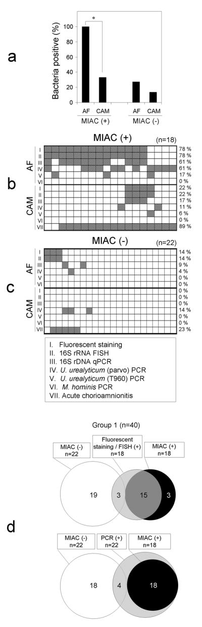
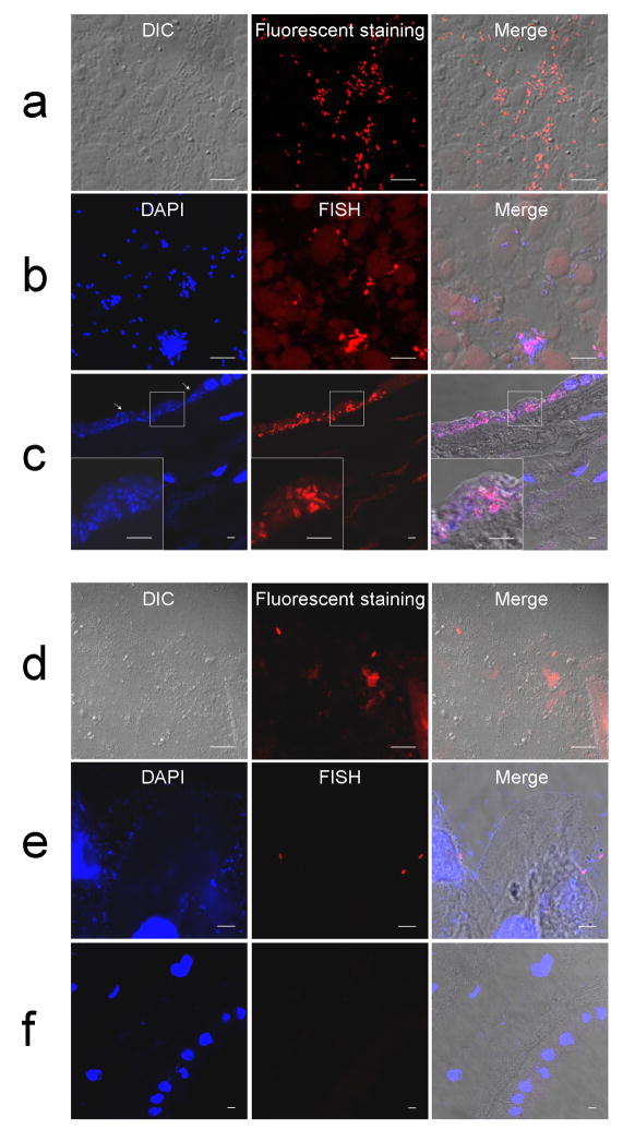
Acute chorioamnionitis is a response to amniotic fluid (AF) infection. However, it remains unclear whether substantial bacterial propagation in the chorioamniotic membranes (CAMs) precedes microbial invasion of the amniotic cavity (MIAC), which is inconsistent with characteristic 'amniotropic neutrophil migration' in acute chorioamnionitis. This study was performed to determine whether CAMs have widespread bacterial infection during MIAC and whether bacteria normally colonize CAMs. AF pellets and CAMs from the following groups were studied: group 1, patients with positive (n=18) or negative (n=22) AF cultures; group 2, patients with or without acute chorioamnionitis in which the amnion and chorion were studied separately (n=60); and group 3, patients at term who underwent a cesarean delivery (n=30). SYTO 9/propidium iodide fluorescent staining and fluorescent in situ hybridization for 16S rRNA were performed. Real-time quantitative PCR for 16S rDNA and PCR for genital mycoplasmas were also conducted. Bacteria were more frequently detected in AF than in CAMs of patients with positive AF culture (100 vs. 33%; P<0.0001). Bacteria were detected more frequently in CAMs as the severity of chorioamnionitis increased (P<0.01). The median 16S rRNA gene copy number in the amnion was significantly greater than in the chorion (group 2; P<0.0001). Bacteria were not detected in CAMs or AF in women at term before labor (group 3). A fraction of patients with chorioamnionitis or MIAC did not have bacteria in CAMs. Collectively, the findings herein indicate that MIAC does not follow widespread infection of CAMs, but precedes it. We propose a model of MIAC: the initial stage is intra-amniotic bacterial invasion through a discrete region of the CAMs, followed by intra-amniotic proliferation, and bacterial invasion of CAMs primarily extends from the amniotic fluid. This study emphasizes the importance of assessing the intra-amniotic compartment for diagnosis and treatment of preterm birth.


Conflict of interest statement

Disclosure/Duality of Interest

The authors state no conflict of interest.

Figures

Figure 1
Figure 1
Comparison of Group 1 cases without (n=22) and with (n=18) MIAC defined by positive amniotic fluid culture. (a) Proportion of the cases in which bacterial traits were demonstrated by any fluorescent staining, FISH, 16S rDNA qPCR, or real-time PCR for U. urealyticum or M. hominis in the amniotic fluid and in the chorioamniotic membranes. Bacteria were more frequently detected in the amniotic fluid than in the chorioamniotic membranes for cases with MIAC (*P<0.0001). (b, c) Maps showing proportion of the cases with histologic chorioamnionitis or the cases in which bacteria were detected in amniotic fluid and chorioamniotic membranes according to assay methods employed. (d) Venn diagrams showing proportion of cases in which bacteria were detected by different techniques. White and black circles represent cases without and with MIAC. Microscopic examinations (fluorescent staining/FISH) or PCR (16S rDNA qPCR, or real-time PCR for U. urealyticum or M. hominis) are in gray circles, for which the number represents the cases positive for bacteria in the amniotic fluid or in the chorioamniotic membranes. MIAC: microbial invasion of the amniotic cavity, AF: amniotic fluid, CAM: chorioamniotic membranes.
Figure 2
Figure 2
Distribution patterns of bacteria detected by either bacterial 16S rRNA FISH using a EUB338 probe or fluorescent staining. (a–c) A case showing numerous rods in amniotic fluid pellets by fluorescent staining (a) and by 16S rRNA FISH (b). (c) 16S rRNA FISH of the chorioamniotic membranes showing bacterial invasion into amniotic epithelium. The invasion is largely restricted to amniotic epithelium. Cytopathic changes of amniotic epithelial cells with loss of nuclei are evident (arrows). (d–f) Another case showing scattered bacteria in amniotic fluid pellets by fluorescent staining (d) and 16S rRNA FISH (e) but not in the chorioamniotic membranes (f). Magnification ×630.
Figure 3
Figure 3
Images of bacteria in two cases of multiple gestations demonstrated by SYTO 9/propidium iodide fluorescent staining. There were isolated MIAC and histologic chorioamnionitis restricted to only one of the fetuses in both cases. (a) Gross feature of the placenta and the amniotic fluid of a twin case. Discoloration of amniotic surface is evident in twin A, and there is stark contrast between the amniocentesis samples. (b, c) A cluster of bacteria in amniotic fluid (b) and bacterial invasion of amniotic epithelial cells (c) demonstrated by fluorescent staining. Live bacteria were stained with SYTO 9 (green fluorescence), and dead bacteria were stained with propidium iodide (red fluorescence). Note the lack of bacteria in the chorioamniotic connective tissue indicating bacterial propagation from the amniotic cavity (arrow). (d) The other case of quadruplets also showing a large number of bacteria in the amniotic fluid pellets and invasion of the amnion in the chorioamniotic membranes by those bacteria on fluorescent staining. Magnification ×630. AF: amniotic fluid, CAM: chorioamniotic membranes.
Figure 4
Figure 4
Bacterial abundance and prevalence in the chorioamniotic membranes in cases with varying degrees of histologic chorioamnionitis (stage 0 (none)/1/2/3). (a) Total proportion of bacterial positive cases in the chorioamniotic membranes was significantly associated between histologic severity of chorioamnionitis (stage 0 (none)/1/2/3) and bacterial prevalence (P<0.01). (b) Proportion of the cases in which bacteria were detected by fluorescent staining, qPCR for universal bacterial 16S rDNA, real-time PCR for U. urealyticum two biovar (parvo and T960), or M. hominis using specific primers and probes. (c) 16S rRNA gene copy numbers were evaluated by real-time quantitative PCR. 16S rRNA gene copy numbers were significantly higher in the amnion (Am) than in the chorion (Cho) in all groups with chorioamnionitis (stage 0 (none)/1/2/3) (*P<0.01).
Figure 5
Figure 5
Two models depicting bacterial trafficking into the chorioamniotic membranes during progression of MIAC. (a) A model of MIAC, whose basic paradigm includes stages of widespread bacterial invasion along the chorioamniotic or choriodecidual planes before they enter the amniotic cavity. The findings in the present study are not consistent with model A. (b) A model of MIAC propagation supported by the findings in this study. It involves initial intra-amniotic entry of bacteria through a restricted cervical region, intra-amniotic proliferation, and subsequent adhesion and invasion of intra-amniotic bacteria into the amnion from the amniotic cavity. MIAC: microbial invasion of the amniotic cavity. Arrows indicate major direction of microbial invasion.

References

    1. Yoon BH, Romero R, Moon J, et al. Differences in the fetal interleukin-6 response to microbial invasion of the amniotic cavity between term and preterm gestation. J Matern Fetal Neonatal Med. 2003;13:32–38. - PubMed
    1. Romero R, Yoon BH, Mazor M, et al. A comparative study of the diagnostic performance of amniotic fluid glucose, white blood cell count, interleukin-6, and gram stain in the detection of microbial invasion in patients with preterm premature rupture of membranes. Am J Obstet Gynecol. 1993;169:839–851. - PubMed
    1. Coultrip LL, Lien JM, Gomez R, et al. The value of amniotic fluid interleukin-6 determination in patients with preterm labor and intact membranes in the detection of microbial invasion of the amniotic cavity. Am J Obstet Gynecol. 1994;171:901–911. - PubMed
    1. Yoon BH, Romero R, Park JS, et al. Microbial invasion of the amniotic cavity with Ureaplasma urealyticum is associated with a robust host response in fetal, amniotic, and maternal compartments. Am J Obstet Gynecol. 1998;179:1254–1260. - PubMed
    1. Romero R, Espinoza J, Goncalves LF, et al. The role of inflammation and infection in preterm birth. Semin Reprod Med. 2007;25:21–39. - PMC - PubMed

Publication types

MeSH terms

Substances

LinkOut - more resources