. 2009 Jul 9;460(7252):278-82.
doi: 10.1038/nature08136. Epub 2009 Jun 7.

A mechanism linking extra centrosomes to chromosomal instability

Affiliations

A mechanism linking extra centrosomes to chromosomal instability

Neil J Ganem et al. Nature. .

Abstract

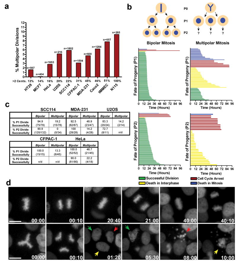
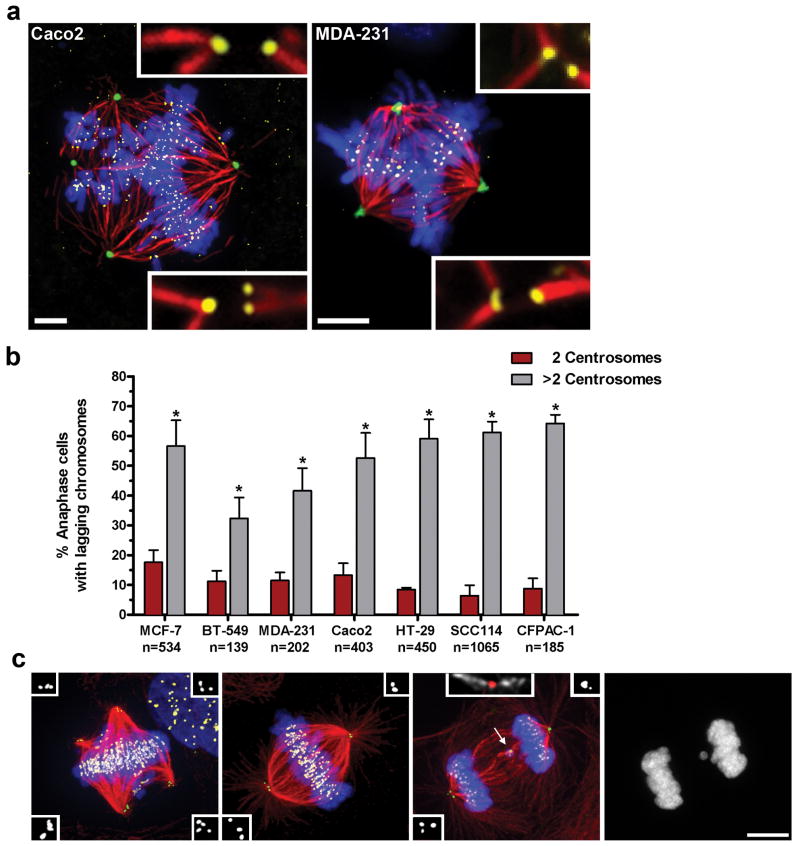
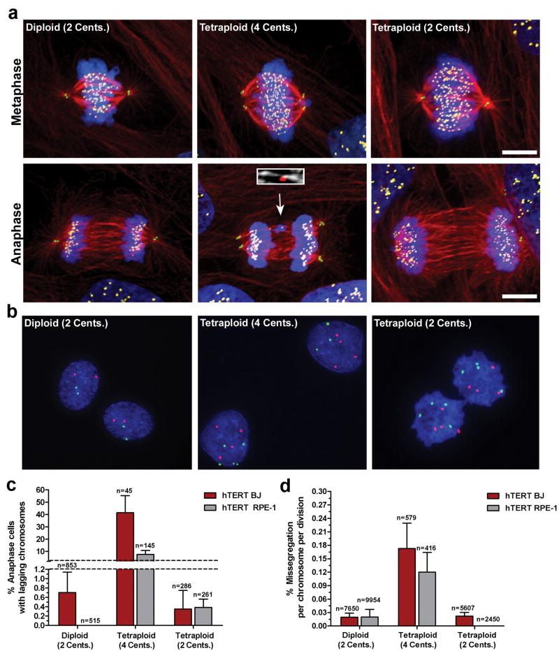
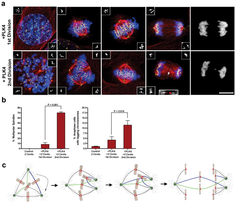
Chromosomal instability (CIN) is a hallmark of many tumours and correlates with the presence of extra centrosomes. However, a direct mechanistic link between extra centrosomes and CIN has not been established. It has been proposed that extra centrosomes generate CIN by promoting multipolar anaphase, a highly abnormal division that produces three or more aneuploid daughter cells. Here we use long-term live-cell imaging to demonstrate that cells with multiple centrosomes rarely undergo multipolar cell divisions, and the progeny of these divisions are typically inviable. Thus, multipolar divisions cannot explain observed rates of CIN. In contrast, we observe that CIN cells with extra centrosomes routinely undergo bipolar cell divisions, but display a significantly increased frequency of lagging chromosomes during anaphase. To define the mechanism underlying this mitotic defect, we generated cells that differ only in their centrosome number. We demonstrate that extra centrosomes alone are sufficient to promote chromosome missegregation during bipolar cell division. These segregation errors are a consequence of cells passing through a transient 'multipolar spindle intermediate' in which merotelic kinetochore-microtubule attachment errors accumulate before centrosome clustering and anaphase. These findings provide a direct mechanistic link between extra centrosomes and CIN, two common characteristics of solid tumours. We propose that this mechanism may be a common underlying cause of CIN in human cancer.


Conflict of interest statement

The authors declare no competing financial interests.

Figures

Figure 1
Figure 1. Multipolar cell divisions are rare and the progeny are typically inviable
a) The percentage of different cancer cells that undergo multipolar cell division based on live-cell imaging (n= number of cell divisions). b) A representative cell fate analysis from SCC114 cells. Individual cell fates of progeny from both bipolar (left column) and multipolar (right column) cell divisions (represented by single colored lines) are shown. c) Percentage of progeny from bipolar and multipolar cell divisions that undergo successful cell division. d) Still frames from the imaging experiment represented in b showing a representative SCC114 cell undergoing several rounds of bipolar cell division (top row), or a single multipolar cell division (bottom row). Colored arrows track the fate of the three progeny from the multipolar cell division (Supp. Movie 1). Time, hours:minutes. Scale bar, 10 μm.
Figure 2
Figure 2. Extra centrosomes correlate with increases in lagging chromosomes
a) Multipolar Caco2 and MDA-231 cells stained for pericentrin (green), microtubules (red), chromosomes (blue), and kinetochores (yellow). Merotelic attachments are shown in insets. Scale bars, 5 μm. b) The percentage of cancer cells with 2 (red) or >2 (grey) centrosomes that exhibit one or more lagging chromosomes during bipolar anaphase (n=number of anaphase cells counted; error bars represent mean ± SE from at least 4 independent experiments; asterisks denote P-values < 0.02 and are derived from an unpaired two-tailed t-test. c) Representative MCF-7 cells with extra centrosomes during prometaphase (multipolar spindle intermediate), metaphase (bipolar spindle with clustered centrosomes), and anaphase (with clustered centrosomes), stained for centrioles (green, and inset white), microtubules (red), chromosomes (blue), and centromeres (yellow). Arrow indicates a lagging chromosome caused by merotelic attachment, in which microtubules emanating from both poles attach to a single kinetochore (inset; microtubules white, centromere red). A detailed description of the criteria used for scoring lagging chromosomes can be found in Supp. Fig. 7. Scale bar, 10 μm.
Figure 3
Figure 3. Extra centrosomes promote chromosome missegregation
a) Human hTERT BJ fibroblasts (Diploid, 2 centrosomes; Tetraploid, 4 centrosomes; Tetraploid, 2 centrosomes) during metaphase and anaphase stained for centrioles (green), microtubules (red), chromosomes (blue), and centromeres (yellow). Arrow indicates a lagging chromosome caused by merotelic attachment (inset; microtubules white, centromere red). b) FISH using centromeric probes specific for chromosomes 6 (green) and 8 (red) in hTERT-BJ fibroblasts. c) Percentage of hTERT BJ (red) and hTERT RPE-1 (grey) cells that exhibit one or more lagging chromosomes during bipolar anaphase (n=number of anaphases counted). d) Missegregation frequency per chromosome per division in hTERT BJ (red) and hTERT RPE-1 (grey) cells (n=number of cell divisions counted). Error bars represent mean ± SE from at least 4 independent experiments. Scale bar, 10 μm.
Figure 4
Figure 4. ‘Multipolar spindle intermediates’ promote merotelic attachment
a) U2OS cells undergoing their 1st or 2nd mitotic division following PLK4 overexpression stained for centrioles (green, inset white), microtubules (red), chromosomes (blue), and centromeres (yellow). Arrow indicates a lagging chromosome caused by merotelic attachment (inset; microtubules white, centromere red). b) The percentage of mitotic cells exhibiting multipolar spindles or lagging chromosomes for each condition. Error bars represent the mean ± SE from 5 independent experiments; P-value derived from paired two-tailed t-test. Scale Bar, 10 μm. c) Extra centrosomes promote merotelic attachment (green microtubules) by altering spindle geometry. In addition, syntelic attachments (blue microtubules) also accumulate upon centrosome clustering and may promote further enhancement of merotely. Unresolved merotelic attachments can give rise to lagging chromosomes at anaphase.

Similar articles

Cited by

References

    1. Lengauer C, Kinzler KW, Vogelstein B. Genetic instability in colorectal cancers. Nature. 1997;386:623–7. - PubMed
    1. D'Assoro AB, Lingle WL, Salisbury JL. Centrosome amplification and the development of cancer. Oncogene. 2002;21:6146–53. - PubMed
    1. Nigg EA. Centrosome aberrations: cause or consequence of cancer progression? Nat Rev Cancer. 2002;2:815–25. - PubMed
    1. Sluder G, Nordberg JJ. The good, the bad and the ugly: the practical consequences of centrosome amplification. Curr Opin Cell Biol. 2004;16:49–54. - PubMed
    1. Rajagopalan H, Lengauer C. Aneuploidy and cancer. Nature. 2004;432:338–41. - PubMed

Publication types