

Using electrical and optical tweezers to facilitate studies of molecular motors
- PMID: 19506758
- PMCID: PMC3639145
- DOI: 10.1039/b821861g
Using electrical and optical tweezers to facilitate studies of molecular motors
Abstract
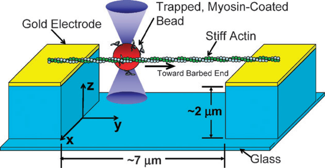
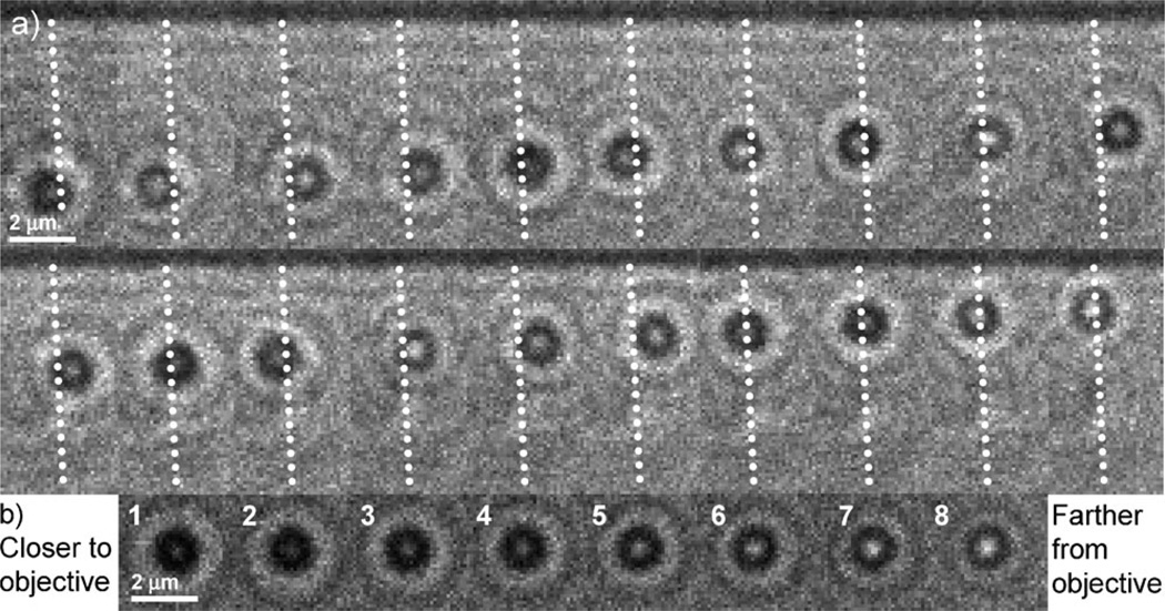
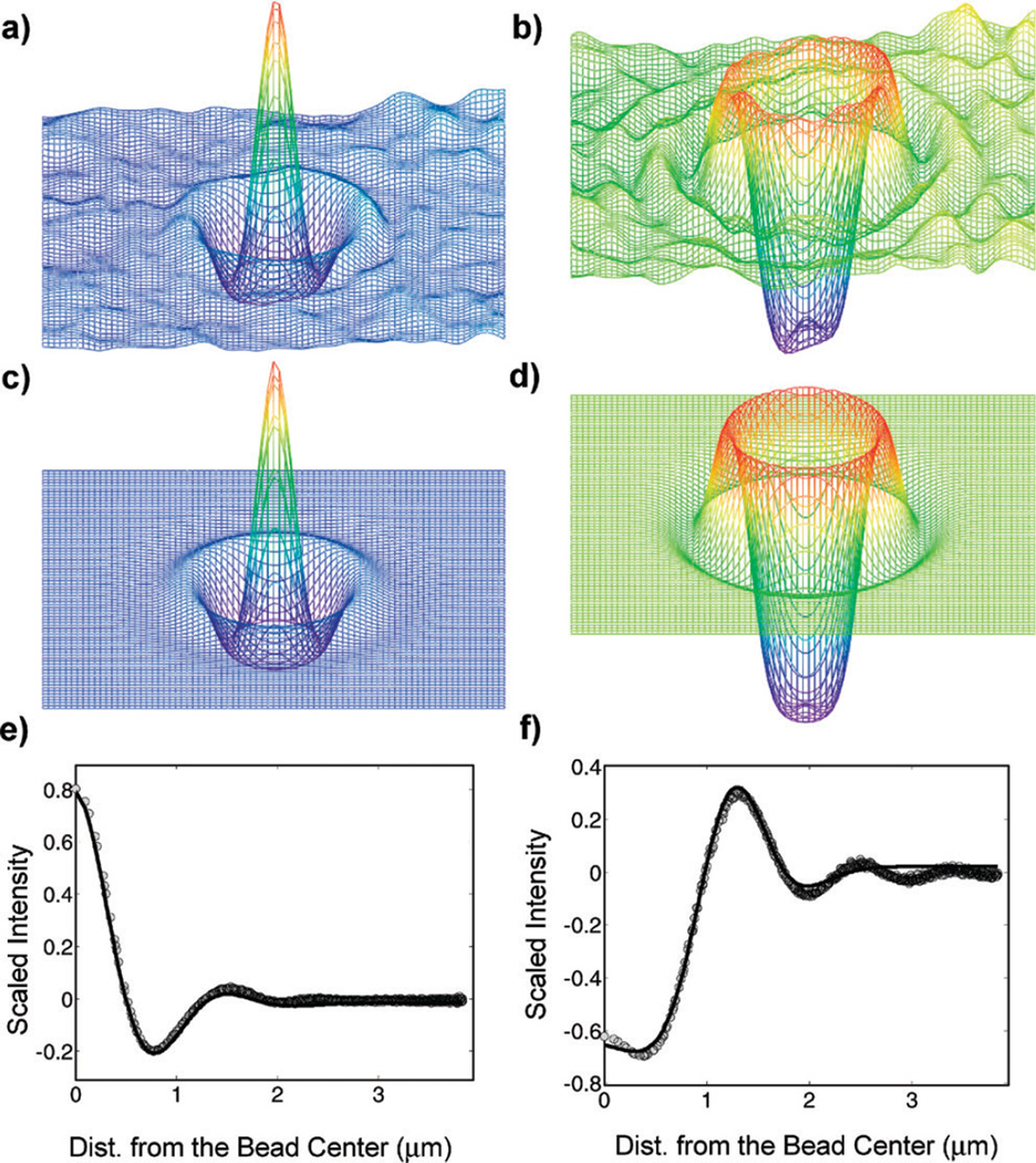
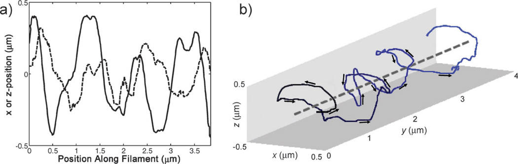
Dielectrophoresis was used to stretch and suspend actin filaments across a trench etched between two electrodes patterned on a glass slide. Optical tweezers were used to bring a motor protein-coated bead into close proximity to a pre-selected, suspended actin filament, facilitating the attachment of the myosin-coated bead to the filament. The clearance beneath the filament allowed the bead to move freely along and around its filamentous track, unhindered by solid surfaces. Using defocused images, the three-dimensional position of the bead was tracked as a function of time to obtain its trajectory. Experiments were carried out with myosin V and myosin X. Both motor proteins followed left-handed helical paths with the myosin X motor exhibiting a shorter pitch than the myosin V. The combined use of electrostatic and optical tweezers facilitates the preparation of motility assays with suspended tracks. Variants of this technique will enable higher complexity experiments in vitro to better understand the behavior of motors in cells.
Figures
References
-
- Schliwa M. Molecular Motors. Weinheim: Wiley-VCH; 2003.
-
- Vale RD. The molecular motor toolbox for intracellular transport. Cell. 2003;112:467–480. - PubMed
-
- Hasson T, Mooseker MS. The growing family of myosin motors and their role in neurons and sensory cells. Curr. Opin. Neurobiol. 1997;7:615–623. - PubMed
-
- Hirokawa N, Takemura R. Molecular motors in neuronal development, intracellular transport and diseases. Curr. Opin. Neurobiol. 2004;14:564–573. - PubMed
-
- Finer JT, Simmons RM, Spudich JA. Single myosin molecule mechanics—piconewton forces and nanometer steps. Nature. 1994;368:113–119. - PubMed
Publication types
MeSH terms
Substances
Grants and funding
LinkOut - more resources
Full Text Sources