

Thymopoiesis in elderly human is associated with systemic inflammatory status
- PMID: 19507053
- PMCID: PMC2693727
- DOI: 10.1007/s11357-008-9084-x
Thymopoiesis in elderly human is associated with systemic inflammatory status
Abstract
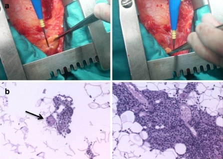
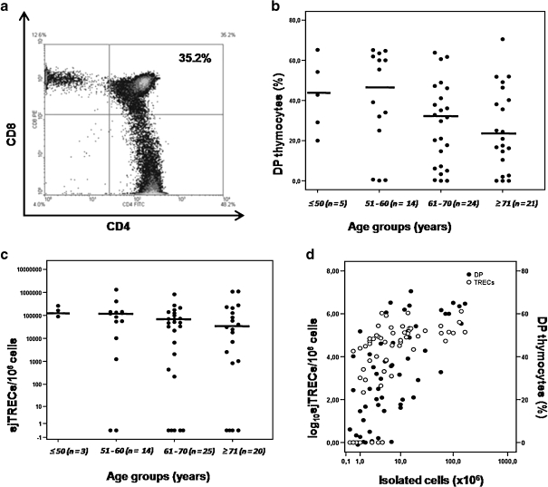
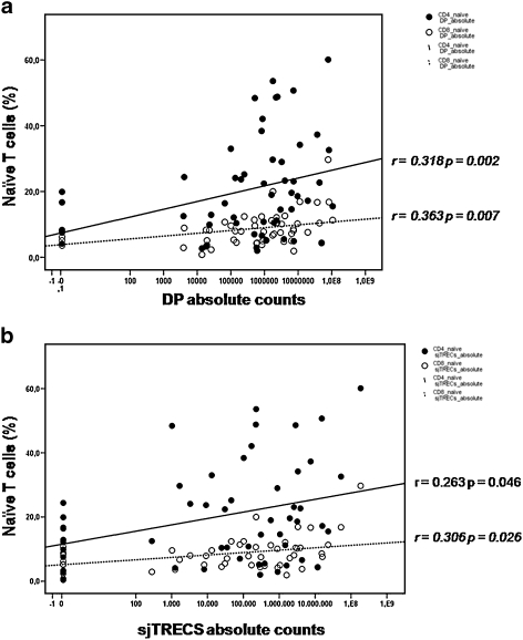
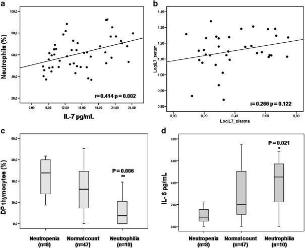
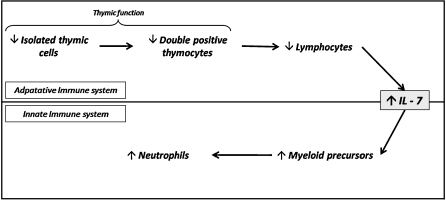
Immunosenescence studies of age-related immune system damage focused on clinical lymphopenic situations or androgenic blockade have revealed new insights about adult human immune reconstitution. However, as far as we know, the extent of lymphopoiesis in the thymus of elderly humans remains unclear. To this effect, we have analyzed 65 adult human thymuses (from 36 to 81 years; median age 68.6 years) obtained from patients who underwent cardiac surgery. Our results show a correlation between CD4(+)CD8(+) double-positive (DP) cells and both the age (inverse) and percentage (direct) of peripheral naive T cells, indicating that the thymus is still able to affect the peripheral lymphocyte pool even in the elderly. We also found significant correlation between the degree of thymopoiesis and the inflammation markers, as shown by the inverse correlations between DP and the percentage of neutrophils and IL-6 levels and the percentage of peripheral lymphocytes. Furthermore, in a multivariate linear regression the percentage of DP and IL-7 levels, but not age, were independently associated with the percentage of neutrophils. In conclusion, the thymus maintains, even in the elderly, an active thymopoiesis that rejuvenates the peripheral naive T-cell pool. Moreover, age-related thymopoietic decay is associated with the peripheral inflammation markers.
Figures
References
-
- {'text': '', 'ref_index': 1, 'ids': [{'type': 'PubMed', 'value': '17237404', 'is_inner': True, 'url': 'https://pubmed.ncbi.nlm.nih.gov/17237404/'}]}
- Aiello FB, Keller JR, Klarmann KD et al (2007) IL-7 induces myelopoiesis and erythropoiesis. J Immunol 178(3):1553–1563 - PubMed
-
- {'text': '', 'ref_index': 1, 'ids': [{'type': 'DOI', 'value': '10.1186/1742-4933-4-9', 'is_inner': False, 'url': 'https://doi.org/10.1186/1742-4933-4-9'}, {'type': 'PMC', 'value': 'PMC2235886', 'is_inner': False, 'url': 'https://pmc.ncbi.nlm.nih.gov/articles/PMC2235886/'}, {'type': 'PubMed', 'value': '18072962', 'is_inner': True, 'url': 'https://pubmed.ncbi.nlm.nih.gov/18072962/'}]}
- Aspinall R, Del Giudice G, Effros RB et al (2007) Challenges for vaccination in the elderly. Immun Ageing 4(1):9. doi:10.1186/1742-4933-4-9 - PMC - PubMed
-
- {'text': '', 'ref_index': 1, 'ids': [{'type': 'DOI', 'value': '10.1111/j.1365-2567.2007.02555.x', 'is_inner': False, 'url': 'https://doi.org/10.1111/j.1365-2567.2007.02555.x'}, {'type': 'PMC', 'value': 'PMC2265901', 'is_inner': False, 'url': 'https://pmc.ncbi.nlm.nih.gov/articles/PMC2265901/'}, {'type': 'PubMed', 'value': '17313487', 'is_inner': True, 'url': 'https://pubmed.ncbi.nlm.nih.gov/17313487/'}]}
- Aw D, Silva AB, Palmer DB et al (2007) Immunosenescence: emerging challenges for an ageing population. Immunology 120(4):435–446. doi:10.1111/j.1365-2567.2007.02555.x - PMC - PubMed
-
- {'text': '', 'ref_index': 1, 'ids': [{'type': 'DOI', 'value': '10.1001/jama.265.4.472', 'is_inner': False, 'url': 'https://doi.org/10.1001/jama.265.4.472'}, {'type': 'PubMed', 'value': '1845912', 'is_inner': True, 'url': 'https://pubmed.ncbi.nlm.nih.gov/1845912/'}]}
- Bauer HM, Ting Y, Greer C et al (1991) Genital human papillomavirus infection in female university student as determined by PCR-based method. JAMA 265:472–477. doi:10.1001/jama.265.4.472 - PubMed
-
- {'text': '', 'ref_index': 1, 'ids': [{'type': 'DOI', 'value': '10.1111/j.0105-2896.2005.00276.x', 'is_inner': False, 'url': 'https://doi.org/10.1111/j.0105-2896.2005.00276.x'}, {'type': 'PubMed', 'value': '15882340', 'is_inner': True, 'url': 'https://pubmed.ncbi.nlm.nih.gov/15882340/'}]}
- Cambier J (2005) Immunosenescence: a problem of lymphopoiesis, homeostasis, microenvironment, and signaling. Immunol Rev 205:5–6. doi:10.1111/j.0105-2896.2005.00276.x - PubMed
LinkOut - more resources
Full Text Sources
Research Materials